Li Yajing, Zhu Zhendong, Cui Haixiang, Ding Kexin, Zhao Yong, Ma Xiangping, Adetunji Adedeji Olufemi, Min Lingjiang
College of Animal Science and Technology, Qingdao Agricultural University, Qingdao 266109, China.
State Key Laboratory of Animal Nutrition, Institute of Animal Sciences, Chinese Academy of Agricultural Sciences, Beijing 100193, China.
Animals (Basel). 2022 Nov 3;12(21):3026. doi: 10.3390/ani12213026.
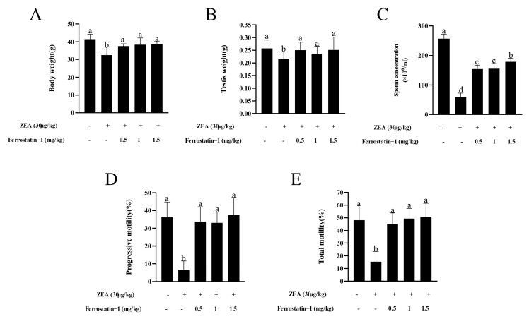
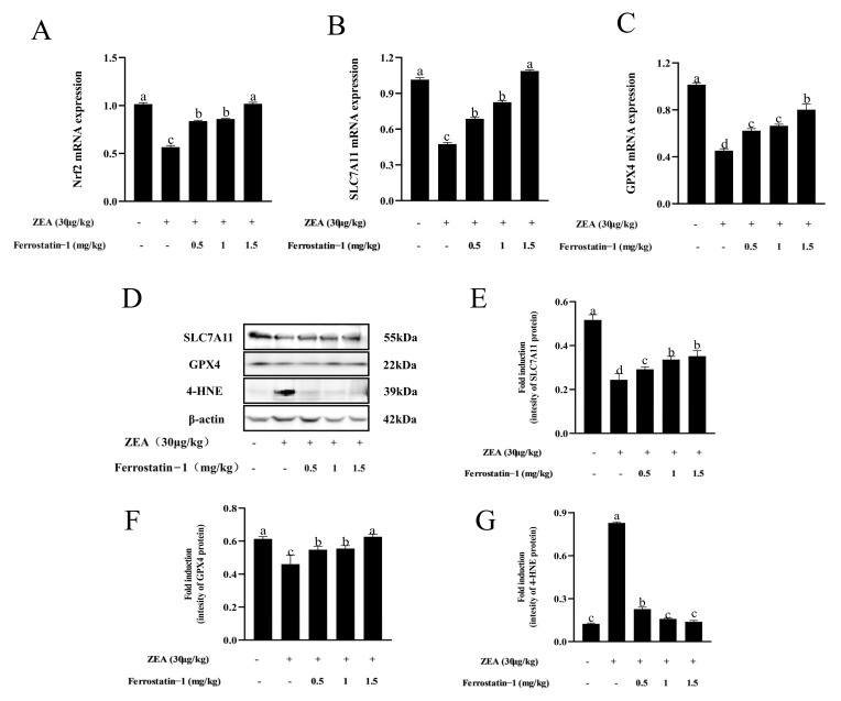
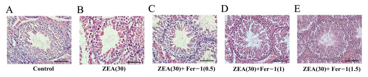
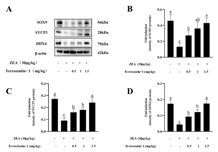
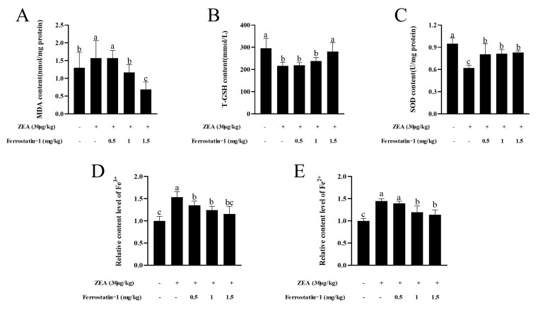
Male reproductive health is critically worsening around the world. It has been reported that the mycotoxin ZEA causes reproductive toxicity to domestic animals and affects spermatogenesis, thereby inhibiting male reproductive function. Ferroptosis is a newly identified type of programmed cell death that is different from apoptosis and it depends on iron accumulation and lipid peroxidation. Whether ferroptosis is linked to ZEA's detrimental effect on spermatogenesis needs to be further explored. This study clarifies ferroptosis's involvement in ZEA-induced damage on spermatogenesis. The reproductive injury model used in this study was induced by gavaging male mice in the ZEA treatment group with 30 μg/kg of ZEA for five weeks. Results show that ZEA treatment reduced mouse sperm motility and concentration, destroyed the structure of the seminiferous tubules of the testis, damaged the antioxidant defense system, and blocked spermatogenesis. Ferrostatin-1 (Fer-1) inhibition of ferroptosis partially alleviated ZEA-induced oligozoospermia in mice. In addition, ZEA treatment was found to activate a signaling pathway associated with ferroptosis in mouse testis. ZEA also downregulated the expression of , , and , and decreased the protein expression of SLC7A11 and GPX4, resulting in the accumulation of lipid peroxides and an increase in the level of 4-HNE protein in the testis. Importantly, these changes were accompanied by an increase in the relative contents of Fe and Fe. Iron accumulation and lipid peroxidation are the causes of ferroptosis in spermatogenic cells, leading to a decrease in sperm motility and concentration. While the administration of Fer-1 at 0.5 and 1 mg/kg also increased the expression of SLC7A11 and GPX4 proteins by upregulating expression, reducing iron accumulation, and reversing ZEA-induced ferroptosis, Fer-1 at 1.5 mg/kg had the best repairing effect for all parameters. In conclusion, ZEA-induced ferroptosis may be mediated by a notable reduction in , and expression levels. Overall, ferroptosis is a novel therapeutic target for mitigating ZEA-induced reproductive toxicity.
全球男性生殖健康状况正在急剧恶化。据报道,霉菌毒素玉米赤霉烯酮(ZEA)会对家畜造成生殖毒性,并影响精子发生,从而抑制男性生殖功能。铁死亡是一种新发现的程序性细胞死亡类型,与凋亡不同,它依赖于铁的积累和脂质过氧化。铁死亡是否与ZEA对精子发生的有害影响有关,还有待进一步探索。本研究阐明了铁死亡在ZEA诱导的精子发生损伤中的作用。本研究中使用的生殖损伤模型是通过给ZEA处理组的雄性小鼠灌胃30μg/kg的ZEA,持续五周诱导而成。结果表明,ZEA处理降低了小鼠精子活力和浓度,破坏了睾丸生精小管的结构,损害了抗氧化防御系统,并阻断了精子发生。铁抑素-1(Fer-1)抑制铁死亡部分缓解了ZEA诱导的小鼠少精子症。此外,发现ZEA处理激活了小鼠睾丸中与铁死亡相关的信号通路。ZEA还下调了……、……和……的表达,并降低了溶质载体家族7成员11(SLC7A11)和谷胱甘肽过氧化物酶4(GPX4)的蛋白表达,导致脂质过氧化物积累和睾丸中4-羟基壬烯醛(4-HNE)蛋白水平升高。重要的是,这些变化伴随着铁(Fe)和亚铁(Fe)相对含量的增加。铁积累和脂质过氧化是生精细胞中铁死亡的原因,导致精子活力和浓度降低。虽然以0.5mg/kg和1mg/kg的剂量施用Fer-1也通过上调……表达、减少铁积累和逆转ZEA诱导的铁死亡来增加SLC7A11和GPX4蛋白的表达,但1.5mg/kg的Fer-1对所有参数的修复效果最佳。总之,ZEA诱导的铁死亡可能是由……、……和……表达水平的显著降低介导的。总体而言,铁死亡是减轻ZEA诱导的生殖毒性的一个新的治疗靶点。