• 文献检索
  • 文档翻译
  • 深度研究
  • 学术资讯
  • Suppr Zotero 插件Zotero 插件
  • 邀请有礼
  • 套餐&价格
  • 历史记录
应用&插件
Suppr Zotero 插件Zotero 插件浏览器插件Mac 客户端Windows 客户端微信小程序
定价
高级版会员购买积分包购买API积分包
服务
文献检索文档翻译深度研究API 文档MCP 服务
关于我们
关于 Suppr公司介绍联系我们用户协议隐私条款
关注我们

Suppr 超能文献

核心技术专利:CN118964589B侵权必究
粤ICP备2023148730 号-1Suppr @ 2026

文献检索

告别复杂PubMed语法,用中文像聊天一样搜索,搜遍4000万医学文献。AI智能推荐,让科研检索更轻松。

立即免费搜索

文件翻译

保留排版,准确专业,支持PDF/Word/PPT等文件格式,支持 12+语言互译。

免费翻译文档

深度研究

AI帮你快速写综述,25分钟生成高质量综述,智能提取关键信息,辅助科研写作。

立即免费体验

基于嵌入式的增强去噪能力的静脉图像处理系统的设计与实现。

Design and Implementation of Embedded-Based Vein Image Processing System with Enhanced Denoising Capabilities.

机构信息

Department of Computer Engineering, Gachon University Global Campus, Seongnam 13120, Korea.

出版信息

Sensors (Basel). 2022 Nov 7;22(21):8559. doi: 10.3390/s22218559.

DOI:10.3390/s22218559
PMID:36366256
原文链接:https://pmc.ncbi.nlm.nih.gov/articles/PMC9656323/
Abstract

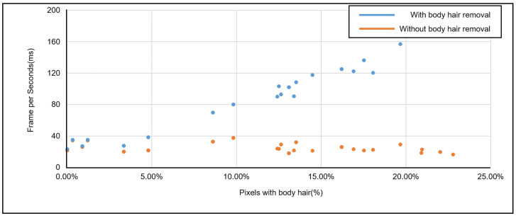
In general, it is very difficult to visually locate blood vessels for intravenous injection or surgery. In addition, if vein detection fails, physical and mental pain occurs to the patient and leads to financial loss in the hospital. In order to prevent this problem, NIR-based vein detection technology is developing. The proposed study combines vein detection and digital hair removal to eliminate body hair, a noise that hinders the accuracy of detection, improving the performance of the entire algorithm by about 10.38% over existing systems. In addition, as a result of performing venous detection of patients without body hair, 5.04% higher performance than the existing system was detected, and the proposed study results were verified. It is expected that the use of devices to which the proposed study is applied will provide more accurate vascular maps in general situations.

摘要

一般来说,很难通过视觉定位静脉进行静脉注射或手术。此外,如果静脉检测失败,患者会感到身心疼痛,并导致医院的经济损失。为了防止这个问题,基于近红外光的静脉检测技术正在发展。本研究将静脉检测与数字脱毛相结合,以消除干扰检测准确性的体毛,从而使整个算法的性能比现有系统提高约 10.38%。此外,由于对没有体毛的患者进行了静脉检测,检测到的性能比现有系统高出 5.04%,验证了本研究的结果。预计应用本研究的设备将在一般情况下提供更准确的血管图。

https://cdn.ncbi.nlm.nih.gov/pmc/blobs/bcea/9656323/52deeca9adc8/sensors-22-08559-g021.jpg
https://cdn.ncbi.nlm.nih.gov/pmc/blobs/bcea/9656323/1955db3ae065/sensors-22-08559-g001.jpg
https://cdn.ncbi.nlm.nih.gov/pmc/blobs/bcea/9656323/ec2567de730b/sensors-22-08559-g002.jpg
https://cdn.ncbi.nlm.nih.gov/pmc/blobs/bcea/9656323/44218e070adb/sensors-22-08559-g003.jpg
https://cdn.ncbi.nlm.nih.gov/pmc/blobs/bcea/9656323/23bac9755767/sensors-22-08559-g004.jpg
https://cdn.ncbi.nlm.nih.gov/pmc/blobs/bcea/9656323/11da65f81471/sensors-22-08559-g005.jpg
https://cdn.ncbi.nlm.nih.gov/pmc/blobs/bcea/9656323/776069eb7f11/sensors-22-08559-g006.jpg
https://cdn.ncbi.nlm.nih.gov/pmc/blobs/bcea/9656323/f110dbf0fc04/sensors-22-08559-g007.jpg
https://cdn.ncbi.nlm.nih.gov/pmc/blobs/bcea/9656323/7607fce9e17a/sensors-22-08559-g008.jpg
https://cdn.ncbi.nlm.nih.gov/pmc/blobs/bcea/9656323/a444939879d8/sensors-22-08559-g009.jpg
https://cdn.ncbi.nlm.nih.gov/pmc/blobs/bcea/9656323/1e025fbbfeeb/sensors-22-08559-g010.jpg
https://cdn.ncbi.nlm.nih.gov/pmc/blobs/bcea/9656323/2e0831a8b696/sensors-22-08559-g011.jpg
https://cdn.ncbi.nlm.nih.gov/pmc/blobs/bcea/9656323/7749b7caff17/sensors-22-08559-g012.jpg
https://cdn.ncbi.nlm.nih.gov/pmc/blobs/bcea/9656323/e981b550a405/sensors-22-08559-g013.jpg
https://cdn.ncbi.nlm.nih.gov/pmc/blobs/bcea/9656323/ae7de8b98f41/sensors-22-08559-g014.jpg
https://cdn.ncbi.nlm.nih.gov/pmc/blobs/bcea/9656323/0d91fd1a7d56/sensors-22-08559-g015.jpg
https://cdn.ncbi.nlm.nih.gov/pmc/blobs/bcea/9656323/3c06c347974e/sensors-22-08559-g016.jpg
https://cdn.ncbi.nlm.nih.gov/pmc/blobs/bcea/9656323/4dd67fe6a80a/sensors-22-08559-g017.jpg
https://cdn.ncbi.nlm.nih.gov/pmc/blobs/bcea/9656323/5f252c06dc9b/sensors-22-08559-g018.jpg
https://cdn.ncbi.nlm.nih.gov/pmc/blobs/bcea/9656323/5f8aae4bb2ab/sensors-22-08559-g019.jpg
https://cdn.ncbi.nlm.nih.gov/pmc/blobs/bcea/9656323/f222e0d799ee/sensors-22-08559-g020.jpg
https://cdn.ncbi.nlm.nih.gov/pmc/blobs/bcea/9656323/52deeca9adc8/sensors-22-08559-g021.jpg

相似文献

1
Design and Implementation of Embedded-Based Vein Image Processing System with Enhanced Denoising Capabilities.基于嵌入式的增强去噪能力的静脉图像处理系统的设计与实现。
Sensors (Basel). 2022 Nov 7;22(21):8559. doi: 10.3390/s22218559.
2
Wavelet denoising for quantum noise removal in chest digital tomosynthesis.用于胸部数字断层合成中去除量子噪声的小波去噪
Int J Comput Assist Radiol Surg. 2015 Jan;10(1):75-86. doi: 10.1007/s11548-014-1003-2. Epub 2014 Apr 20.
3
Image denoising substantially improves accuracy and precision of intravoxel incoherent motion parameter estimates.图像去噪显著提高了体素内不相干运动参数估计的准确性和精确性。
PLoS One. 2017 Apr 5;12(4):e0175106. doi: 10.1371/journal.pone.0175106. eCollection 2017.
4
Iterative image-domain decomposition for dual-energy CT.双能CT的迭代图像域分解
Med Phys. 2014 Apr;41(4):041901. doi: 10.1118/1.4866386.
5
Combined iterative reconstruction and image-domain decomposition for dual energy CT using total-variation regularization.使用全变差正则化的双能CT的联合迭代重建与图像域分解
Med Phys. 2014 May;41(5):051909. doi: 10.1118/1.4870375.
6
Efficient learning representation of noise-reduced foam effects with convolutional denoising networks.利用卷积去噪网络实现降噪泡沫效果的高效学习表示。
PLoS One. 2022 Oct 10;17(10):e0275117. doi: 10.1371/journal.pone.0275117. eCollection 2022.
7
Oral CT Image Processing Based on Oral CT Image Filtering Algorithm.基于口腔 CT 图像滤波算法的口腔 CT 图像处理。
Comput Intell Neurosci. 2022 Sep 19;2022:6041872. doi: 10.1155/2022/6041872. eCollection 2022.
8
A denoising algorithm for projection measurements in cone-beam computed tomography.一种用于锥束计算机断层摄影投影测量的去噪算法。
Comput Biol Med. 2016 Feb 1;69:71-82. doi: 10.1016/j.compbiomed.2015.12.007. Epub 2015 Dec 19.
9
A Novel Algorithm for Hyperspectral Image Denoising in Medical Application.一种用于医学应用的高光谱图像去噪的新算法。
J Med Syst. 2019 Jul 23;43(9):291. doi: 10.1007/s10916-019-1403-5.
10
Adaptive nonlocal means filtering based on local noise level for CT denoising.基于局部噪声水平的自适应非局部均值滤波用于CT去噪。
Med Phys. 2014 Jan;41(1):011908. doi: 10.1118/1.4851635.

引用本文的文献

1
Antimicrobial photodynamic therapy against Escherichia coli by exploiting endogenously produced Protoporphyrin IX- In vitro study.利用内源性产生的原卟啉 IX 对抗大肠杆菌的光动力抗菌治疗——体外研究。
Lasers Med Sci. 2024 Aug 1;39(1):204. doi: 10.1007/s10103-024-04150-8.
2
Photoacoustic imaging for cutaneous melanoma assessment: a comprehensive review.光声成像在皮肤黑色素瘤评估中的应用:全面综述。
J Biomed Opt. 2024 Jan;29(Suppl 1):S11518. doi: 10.1117/1.JBO.29.S1.S11518. Epub 2024 Jan 12.

本文引用的文献

1
Hair removal in dermoscopy images using variational autoencoders.使用变分自编码器去除皮肤镜图像中的毛发。
Skin Res Technol. 2022 May;28(3):445-454. doi: 10.1111/srt.13145. Epub 2022 Mar 7.
2
Performance Evaluation of Dorsal Vein Network of Hand Imaging Using Relative Total Variation-Based Regularization for Smoothing Technique in a Miniaturized Vein Imaging System: A Pilot Study.应用基于相对全变差正则化的平滑技术对手部背静脉网络成像性能评估在小型静脉成像系统中的应用:初步研究。
Int J Environ Res Public Health. 2021 Feb 6;18(4):1548. doi: 10.3390/ijerph18041548.
3
Digital hair segmentation using hybrid convolutional and recurrent neural networks architecture.
基于混合卷积和循环神经网络架构的数字头发分割。
Comput Methods Programs Biomed. 2019 Aug;177:17-30. doi: 10.1016/j.cmpb.2019.05.010. Epub 2019 May 15.
4
Machine learning approaches in medical image analysis: From detection to diagnosis.机器学习在医学图像分析中的应用:从检测到诊断。
Med Image Anal. 2016 Oct;33:94-97. doi: 10.1016/j.media.2016.06.032. Epub 2016 Jun 23.
5
Augmented reality based real-time subcutaneous vein imaging system.基于增强现实的实时皮下静脉成像系统。
Biomed Opt Express. 2016 Jun 13;7(7):2565-85. doi: 10.1364/BOE.7.002565. eCollection 2016 Jul 1.
6
Utilizing the vein viewer technology to map out a venous flap preoperatively.术前利用静脉显像技术绘制静脉皮瓣。
J Reconstr Microsurg. 2013 Jul;29(6):423-4. doi: 10.1055/s-0033-1343951. Epub 2013 Apr 19.
7
Efficacy of VeinViewer in pediatric peripheral intravenous access: a randomized controlled trial.静脉可视仪在儿科外周静脉穿刺中的效果:一项随机对照试验。
Eur J Pediatr. 2012 Jul;171(7):1121-5. doi: 10.1007/s00431-012-1713-9. Epub 2012 Mar 14.
8
Image quality assessment: from error visibility to structural similarity.图像质量评估:从误差可见性到结构相似性。
IEEE Trans Image Process. 2004 Apr;13(4):600-12. doi: 10.1109/tip.2003.819861.