• 文献检索
  • 文档翻译
  • 深度研究
  • 学术资讯
  • Suppr Zotero 插件Zotero 插件
  • 邀请有礼
  • 套餐&价格
  • 历史记录
应用&插件
Suppr Zotero 插件Zotero 插件浏览器插件Mac 客户端Windows 客户端微信小程序
定价
高级版会员购买积分包购买API积分包
服务
文献检索文档翻译深度研究API 文档MCP 服务
关于我们
关于 Suppr公司介绍联系我们用户协议隐私条款
关注我们

Suppr 超能文献

核心技术专利:CN118964589B侵权必究
粤ICP备2023148730 号-1Suppr @ 2026

文献检索

告别复杂PubMed语法,用中文像聊天一样搜索,搜遍4000万医学文献。AI智能推荐,让科研检索更轻松。

立即免费搜索

文件翻译

保留排版,准确专业,支持PDF/Word/PPT等文件格式,支持 12+语言互译。

免费翻译文档

深度研究

AI帮你快速写综述,25分钟生成高质量综述,智能提取关键信息,辅助科研写作。

立即免费体验

文莱人群上颌第一和第二磨牙近中颊根第二根管的患病率——锥形束CT分析

Prevalence of mesiobuccal-2 canals in maxillary first and second molars among the Bruneian population-CBCT analysis.

作者信息

Onn Hui Yi, Sikun Malissa Siao Yun Abdullah, Abdul Rahman Hanif, Dhaliwal Jagjit Singh

机构信息

PAPRSB Institute of Health Sciences, Universiti Brunei Darussalam, Gadong, Bandar Seri Begawan, BE1410, Brunei Darussalam.

Ministry of Health, Department of Dental Services, Bandar Seri Begawan, Brunei Darussalam.

出版信息

BDJ Open. 2022 Nov 19;8(1):32. doi: 10.1038/s41405-022-00125-5.

DOI:10.1038/s41405-022-00125-5
PMID:36402745
原文链接:https://pmc.ncbi.nlm.nih.gov/articles/PMC9675843/
Abstract

INTRODUCTION

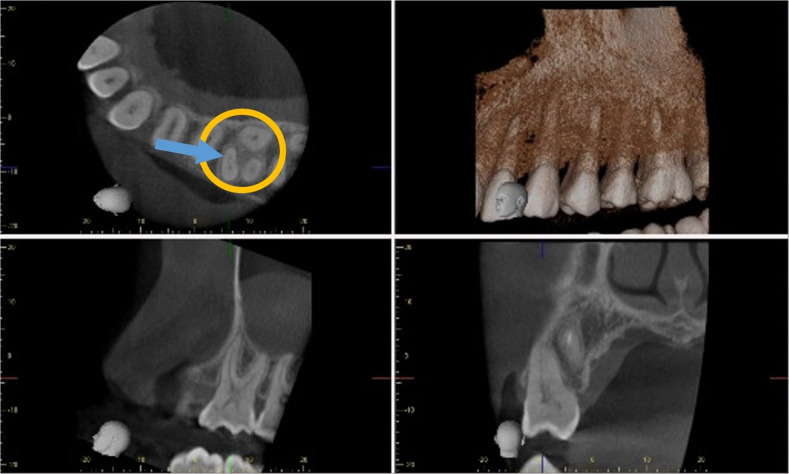
Identification of the second mesiobuccal canal (MB-2) in maxillary molars is considered an endodontic concern of many practitioners due to its complex morphology. The use of Cone-beam Computed Tomography (CBCT) is a necessity for easier location of this elusive canal during endodontic treatment.

AIM

To study the prevalence of the MB-2 canal in the maxillary first and second molars amongst the Bruneian population.

MATERIALS AND METHODS

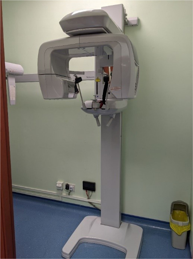
A retrospective study involving a review of scans taken from a CBCT scanner (J Morita; Veraviewepocs 3D R100 Panoramic/Cephalometric) over a 5-year period, from May 2016 to May 2021 was carried out. A total of 342 maxillary molars were evaluated independently by two observers. Any contradicting outcomes were discussed by both observers until a consensus was reached. In addition, the correlation of MB-2 canals with gender and age were calculated using the chi-squared test.

RESULTS

The prevalence of MB-2 canal in the maxillary first and second molars are 51.3% and 29.8% respectively. Both males and females have a similar prevalence of MB-2 canals in the maxillary first and second molars. The incidence of MB-2 canals in both maxillary first and second molars significantly decreases with increasing age. No significant correlation between the prevalence of MB-2 canals with different gender groups in the population.

CONCLUSIONS

It is crucial for clinicians to identify the presence of MB-2 canals when performing endodontic treatment of the maxillary first and second molars. Varying prevalence has been reported for different populations. Recognising this wide-ranging prevalence amongst different populations will allow for greater predictability in ensuring endodontic treatment success.

摘要

引言

由于上颌磨牙的第二近中颊根管(MB-2)形态复杂,其识别被许多从业者视为牙髓治疗中的一个关注点。在牙髓治疗过程中,使用锥形束计算机断层扫描(CBCT)对于更容易定位这条难以捉摸的根管是必要的。

目的

研究文莱人群上颌第一和第二磨牙中MB-2根管的发生率。

材料与方法

进行了一项回顾性研究,回顾了2016年5月至2021年5月期间使用CBCT扫描仪(J Morita;Veraviewepocs 3D R100全景/头影测量仪)进行的扫描。两位观察者独立评估了总共342颗上颌磨牙。任何有矛盾的结果都由两位观察者进行讨论,直到达成共识。此外,使用卡方检验计算MB-2根管与性别和年龄的相关性。

结果

上颌第一和第二磨牙中MB-2根管的发生率分别为51.3%和29.8%。男性和女性在上颌第一和第二磨牙中MB-2根管的发生率相似。上颌第一和第二磨牙中MB-2根管的发生率均随年龄增长而显著降低。人群中不同性别组的MB-2根管发生率之间无显著相关性。

结论

临床医生在对上颌第一和第二磨牙进行牙髓治疗时识别MB-2根管的存在至关重要。不同人群的发生率有所不同。认识到不同人群中这种广泛的发生率将有助于在确保牙髓治疗成功方面提高可预测性。

https://cdn.ncbi.nlm.nih.gov/pmc/blobs/5ba2/9675843/596ede41107c/41405_2022_125_Fig3_HTML.jpg
https://cdn.ncbi.nlm.nih.gov/pmc/blobs/5ba2/9675843/b5dbbf3e4abd/41405_2022_125_Fig1_HTML.jpg
https://cdn.ncbi.nlm.nih.gov/pmc/blobs/5ba2/9675843/3312779b86c9/41405_2022_125_Fig2_HTML.jpg
https://cdn.ncbi.nlm.nih.gov/pmc/blobs/5ba2/9675843/596ede41107c/41405_2022_125_Fig3_HTML.jpg
https://cdn.ncbi.nlm.nih.gov/pmc/blobs/5ba2/9675843/b5dbbf3e4abd/41405_2022_125_Fig1_HTML.jpg
https://cdn.ncbi.nlm.nih.gov/pmc/blobs/5ba2/9675843/3312779b86c9/41405_2022_125_Fig2_HTML.jpg
https://cdn.ncbi.nlm.nih.gov/pmc/blobs/5ba2/9675843/596ede41107c/41405_2022_125_Fig3_HTML.jpg

相似文献

1
Prevalence of mesiobuccal-2 canals in maxillary first and second molars among the Bruneian population-CBCT analysis.文莱人群上颌第一和第二磨牙近中颊根第二根管的患病率——锥形束CT分析
BDJ Open. 2022 Nov 19;8(1):32. doi: 10.1038/s41405-022-00125-5.
2
A Cone-beam Computed Tomographic Study of Root and Canal Morphology of Maxillary First and Second Permanent Molars in a Thai Population.一项泰国人群上颌第一和第二恒磨牙根管及形态的锥形束 CT 研究。
J Endod. 2018 Jan;44(1):56-61. doi: 10.1016/j.joen.2017.08.020. Epub 2017 Oct 20.
3
The morphology of maxillary first and second molars analyzed by cone-beam computed tomography in a polish population.通过锥形束计算机断层扫描对波兰人群上颌第一和第二磨牙形态进行的分析。
BMC Med Imaging. 2017 Dec 29;17(1):68. doi: 10.1186/s12880-017-0243-3.
4
The Incidence of Second Mesiobuccal Canals Located in Maxillary Molars with the Aid of Cone-beam Computed Tomography.锥形束计算机断层扫描辅助下上颌磨牙第二近中颊管的发生率。
J Endod. 2018 Apr;44(4):565-570. doi: 10.1016/j.joen.2017.08.026. Epub 2017 Nov 15.
5
Assessment of the presence of a second mesiobuccal canal in maxillary first molars according to the location of the main mesiobuccal canal-a micro-computed tomographic study.上颌第一磨牙第二近中颊根管存在情况的评估:基于主近中颊根管位置的分析-一项显微 CT 研究。
Clin Oral Investig. 2021 Jun;25(6):3937-3944. doi: 10.1007/s00784-020-03723-5. Epub 2021 Jan 6.
6
Root Canal Morphologies of Mesiobuccal Roots of Maxillary Molars using Cone beam Computed Tomography and Periapical Radiographic Techniques in an Iranian Population.使用锥形束计算机断层扫描和根尖X线摄影技术对伊朗人群上颌磨牙近中颊根根管形态的研究
J Contemp Dent Pract. 2017 Sep 1;18(9):745-749. doi: 10.5005/jp-journals-10024-2119.
7
Detection of Second Mesiobuccal Canals in Maxillary First Molars Using a New Angle of Cone Beam Computed Tomography.使用锥束计算机断层扫描新角度检测上颌第一磨牙的第二近中颊根管
Iran J Radiol. 2016 Jun 29;13(4):e31155. doi: 10.5812/iranjradiol.31155. eCollection 2016 Oct.
8
Ex vivo detection of mesiobuccal canals in maxillary molars using CBCT at four different isotropic voxel dimensions.使用四种不同各向同性体素尺寸的 CBCT 在下颌磨牙的 ex vivo 检测中检测近中颊管。
Int Endod J. 2011 Aug;44(8):752-8. doi: 10.1111/j.1365-2591.2011.01882.x. Epub 2011 Apr 7.
9
Evaluation of Distolingual Canal/Roots in Mandibular Molars and Mesiobuccal Canals in Maxillary Molars by CBCT.CBCT对下颌磨牙远中舌侧根管/牙根及上颌磨牙近中颊侧根管的评估
Open Dent J. 2017 Jun 30;11:360-366. doi: 10.2174/1874210601711010360. eCollection 2017.
10
Root Morphology of the Maxillary First and Second Molars in an Iranian Population Using Cone Beam Computed Tomography.使用锥形束计算机断层扫描技术对上颌第一和第二磨牙在伊朗人群中的牙根形态进行研究。
J Dent (Tehran). 2017 May;14(3):115-122.

引用本文的文献

1
Five-canal maxillary first molar with calcified MB2 and low bifurcation of distal root canals: a case report.伴有钙化的MB2和远中根管低分叉的五根管上颌第一磨牙:一例报告
Front Oral Health. 2025 Mar 28;6:1511699. doi: 10.3389/froh.2025.1511699. eCollection 2025.
2
Machine Learning Models in the Detection of MB2 Canal Orifice in CBCT Images.机器学习模型在锥束计算机断层扫描(CBCT)图像中检测MB2根管口的应用
Int Dent J. 2025 Jun;75(3):1640-1648. doi: 10.1016/j.identj.2025.02.025. Epub 2025 Mar 25.
3
Two Palatal Roots in Maxillary First Molar, a Rare Entity: Report of Two Cases.

本文引用的文献

1
Sample size calculation for prevalence studies using Scalex and ScalaR calculators.使用 Scalex 和 ScalaR 计算器进行流行率研究的样本量计算。
BMC Med Res Methodol. 2022 Jul 30;22(1):209. doi: 10.1186/s12874-022-01694-7.
2
Assessment of Mesiobuccal Canal Configuration, Prevalence and Inter-Orifice Distance at Different Root Thirds of Maxillary First Molars: A CBCT Study.上颌第一磨牙不同牙根三分之一处近中颊根管形态、发生率及根管口间距的评估:一项锥形束CT研究
Clin Cosmet Investig Dent. 2021 Mar 24;13:105-111. doi: 10.2147/CCIDE.S300241. eCollection 2021.
3
The frequency of the second mesiobuccal canal in maxillary first molars among a sample of the Kurdistan Region-Iraq population - A retrospective cone-beam computed tomography evaluation.
上颌第一磨牙双腭根:一种罕见情况——两例报告
J Dent (Shiraz). 2024 Jun 1;25(2):178-182. doi: 10.30476/dentjods.2023.98649.2101. eCollection 2024 Jun.
4
Cone beam computed tomography analysis of the root and canal morphology of the maxillary second molars in a Hail province of the Saudi population.沙特人群中某一省上颌第二磨牙牙根及根管形态的锥束计算机断层扫描分析
Heliyon. 2023 Aug 26;9(9):e19477. doi: 10.1016/j.heliyon.2023.e19477. eCollection 2023 Sep.
伊拉克库尔德地区人群样本中上颌第一磨牙近中颊根第二根管的发生率——一项回顾性锥形束计算机断层扫描评估
J Dent Sci. 2021 Jan;16(1):91-95. doi: 10.1016/j.jds.2020.06.021. Epub 2020 Jul 18.
4
A cone-beam computed tomography study of the prevalence and location of the second mesiobuccal root canal in maxillary molars.一项关于上颌磨牙近中颊侧第二根管患病率及位置的锥形束计算机断层扫描研究。
Restor Dent Endod. 2020 Sep 3;45(4):e46. doi: 10.5395/rde.2020.45.e46. eCollection 2020 Nov.
5
Missed canals in endodontically treated maxillary molars of a Brazilian subpopulation: prevalence and association with periapical lesion using cone-beam computed tomography.巴西亚人群上颌磨牙根管治疗后遗漏根管的情况:使用锥形束 CT 评估其与根尖病变的相关性及患病率。
Clin Oral Investig. 2021 Apr;25(4):2317-2323. doi: 10.1007/s00784-020-03554-4. Epub 2020 Sep 2.
6
Morphometric analysis of panoramic mandibular index, mental index, and antegonial index.下颌全景指数、颏部指数和前颌弓指数的形态分析。
J Int Med Res. 2020 Mar;48(3):300060520912138. doi: 10.1177/0300060520912138.
7
Second mesiobuccal root canal in maxillary molars-A systematic review and meta-analysis of prevalence studies using cone beam computed tomography.上颌磨牙第二近颊根管:锥形束 CT 研究的系统评价和荟萃分析。
Arch Oral Biol. 2020 May;113:104589. doi: 10.1016/j.archoralbio.2019.104589. Epub 2019 Oct 24.
8
The Influence of Missed Canals on the Prevalence of Periapical Lesions in Endodontically Treated Teeth: A Cross-sectional Study.未发现根管对根管治疗后牙齿根尖病变发生率的影响:一项横断面研究。
J Endod. 2020 Jan;46(1):34-39.e1. doi: 10.1016/j.joen.2019.10.007. Epub 2019 Nov 14.
9
The second mesiobuccal canal in three-rooted maxillary first molar of Karnataka Indian sub-populations: A cone-beam computed tomography study.印度卡纳塔克邦亚人群上颌第一磨牙三根中第二近中颊根的锥形束计算机断层扫描研究。
J Oral Biol Craniofac Res. 2019 Oct-Dec;9(4):347-351. doi: 10.1016/j.jobcr.2019.08.001. Epub 2019 Aug 31.
10
The 5-Year Survival Rate of Nonsurgical Endodontic Treatment: A Population-based Cohort Study in Korea.非手术根管治疗的 5 年生存率:韩国一项基于人群的队列研究。
J Endod. 2019 Oct;45(10):1192-1199. doi: 10.1016/j.joen.2019.07.004. Epub 2019 Aug 19.