Radiation Oncology Branch, Center for Cancer Research, National Cancer Institute (NCI), Bethesda, MD, 20892, USA.
Sci Rep. 2022 Nov 19;12(1):19941. doi: 10.1038/s41598-022-24051-6.
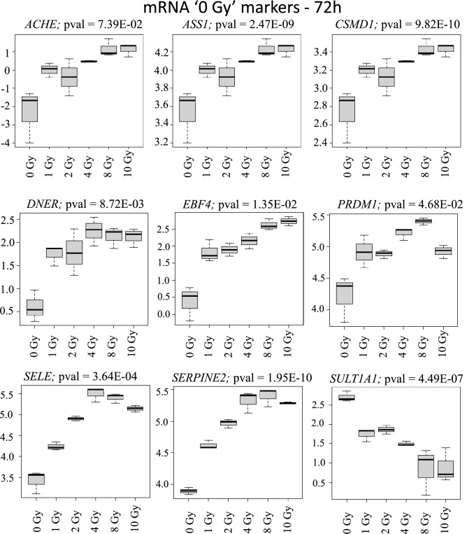
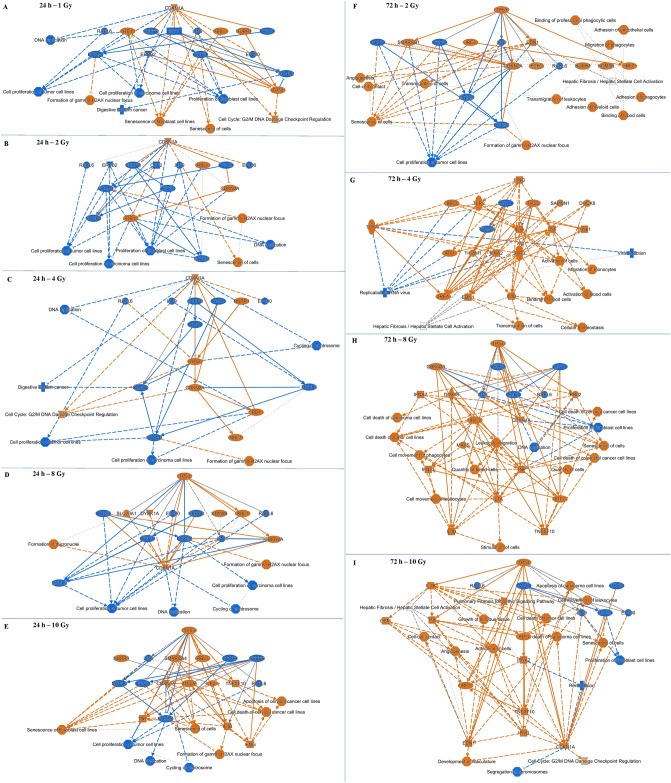
Recent and past research have highlighted the importance of the endothelium in the manifestation of radiation injury. Our primary focus is on medical triage and management following whole body or partial-body irradiation. Here we investigated the usability of endothelial cells' radiation response for biodosimetry applications. We profiled the transcriptome in cultured human endothelial cells treated with increasing doses of X-rays. mRNA expression changes were useful 24 h and 72 h post-radiation, microRNA and lncRNA expression changes were useful 72 h after radiation. More mRNA expressions were repressed than induced while more miRNA and lncRNA expressions were induced than repressed. These novel observations imply distinct radiation responsive regulatory mechanisms for coding and non-coding transcripts. It also follows how different RNA species should be explored as biomarkers for different time-points. Radiation-responsive markers which could classify no radiation (i.e., '0 Gy') and dose-differentiating markers were also predicted. IPA analysis showed growth arrest-related processes at 24 h but immune response coordination at the 72 h post-radiation. Collectively, these observations suggest that endothelial cells have a precise dose and time-dependent response to radiation. Further studies in the laboratory are examining if these differences could be captured in the extracellular vesicles released by irradiated endothelial cells.
最近和过去的研究都强调了内皮细胞在辐射损伤表现中的重要性。我们主要关注全身或半身照射后的医疗分诊和管理。在这里,我们研究了内皮细胞对辐射的反应在生物剂量测定中的可用性。我们对经不同剂量 X 射线处理的培养人内皮细胞进行了转录组分析。mRNA 表达变化在辐射后 24 小时和 72 小时有用,miRNA 和 lncRNA 表达变化在辐射后 72 小时有用。受抑制的 mRNA 表达多于诱导的,而受诱导的 miRNA 和 lncRNA 表达多于受抑制的。这些新的观察结果表明编码和非编码转录物的辐射反应调节机制不同。这也说明了不同的 RNA 种类应该如何作为不同时间点的生物标志物进行探索。还预测了能够区分无辐射(即“0Gy”)和剂量区分标记的辐射反应标记。IPA 分析显示,在 24 小时时有与生长抑制相关的过程,但在辐射后 72 小时时有免疫反应协调。总的来说,这些观察结果表明内皮细胞对辐射有精确的剂量和时间依赖性反应。实验室中的进一步研究正在检查这些差异是否可以在受照射的内皮细胞释放的细胞外囊泡中捕捉到。