Li Yajing, Li Sen, Xue Xiaoyong, Wang Ting, Li Xiaojiaoyang
School of Life Sciences, Beijing University of Chinese Medicine, Beijing 100029, China.
Beijing Research Institute of Chinese Medicine, Beijing University of Chinese Medicine, Beijing 100029, China.
Chin Herb Med. 2022 Sep 27;14(4):563-575. doi: 10.1016/j.chmed.2022.02.006. eCollection 2022 Oct.
glycoside (TG) is widely used in clinical practice for its multiple bioactivities including anti-inflammatory and immunosuppressive effects. However, emerging studies have frequently reported TG-induced adverse reactions to multiple organs, especially liver. Here, this study aimed to investigate the mechanism of liver damage induced by TG and explore representative components to reflect TG hepatotoxicity.
Network pharmacology was used to determine the potential targets of bile duct injury caused by TG. Next, the hepatotoxic effects of TG, triptolide (TP) and celastrol (CEL) were investigated and compared and . Liver function was determined by measuring serum transaminase and histopathology staining. The cell proliferation and apoptosis were determined by cell viability assay, scratch assay and flow cytometry. The expression of gene of interest was determined by qPCR and Western blot.
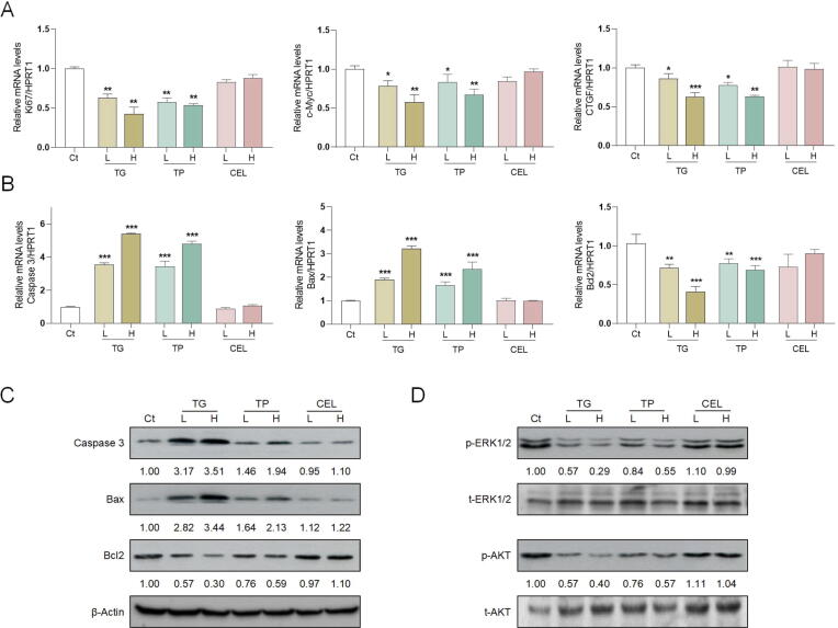
Based on the network pharmacological analysis of 12 bioactive ingredients found in TG, a total of 35 targets and 15 pathways related to bile duct injury were obtained. Both TG and TP resulted in cholangiocyte damage and liver injury, as illustrated by increased levels of serum transaminase and oxidative stress, stimulated portal edema and lymphocytic infiltration and decreased expression of cholangiocyte marker, cytoskeletal 19. In addition, TG and TP inhibited cell proliferation and migration, arrested cell cycle and promoted Caspase-dependent apoptosis of cholangiocytes suppressing the phosphorylation of extracellular regulated protein kinases 1/2 (ERK1/2) and protein kinase B (AKT). While, CEL at equivalent dosage had no obvious hepatotoxicity.
We revealed that TG-stimulated liver injury was specifically characterized by cholangiocyte damage and TP might be the decisive ingredient to reflect TG hepatotoxicity. Our results not only provide novel insights into the mechanism underlying the hepatotoxicity effects of TG but also offer reference for clinical rational use of TG.
雷公藤多苷(TG)因其多种生物活性,包括抗炎和免疫抑制作用,在临床实践中被广泛应用。然而,新出现的研究频繁报道TG对多个器官,尤其是肝脏,会引起不良反应。在此,本研究旨在探讨TG诱导肝损伤的机制,并探索反映TG肝毒性的代表性成分。
采用网络药理学确定TG引起胆管损伤的潜在靶点。接下来,研究并比较了TG、雷公藤甲素(TP)和雷公藤红素(CEL)的肝毒性作用。通过检测血清转氨酶和组织病理学染色来测定肝功能。通过细胞活力测定、划痕试验和流式细胞术来测定细胞增殖和凋亡。通过qPCR和蛋白质免疫印迹法来测定目标基因的表达。
基于对TG中发现的12种生物活性成分的网络药理学分析,共获得了35个靶点和15条与胆管损伤相关的通路。TG和TP均导致胆管细胞损伤和肝损伤,血清转氨酶水平升高和氧化应激增加、门静脉水肿和淋巴细胞浸润以及胆管细胞标志物细胞角蛋白19表达降低均说明了这一点。此外,TG和TP抑制细胞增殖和迁移,使细胞周期停滞,并促进胆管细胞的Caspase依赖性凋亡,同时抑制细胞外调节蛋白激酶1/2(ERK1/2)和蛋白激酶B(AKT)的磷酸化。而同等剂量的CEL没有明显的肝毒性。
我们发现TG刺激引起的肝损伤具有胆管细胞损伤的特异性特征,TP可能是反映TG肝毒性的决定性成分。我们的研究结果不仅为TG肝毒性作用的潜在机制提供了新的见解,也为TG的临床合理使用提供了参考。