Department of Neurology and Neurological Science, Graduate School of Medical and Dental Science, Tokyo Medical and Dental University, Tokyo, Japan.
Radiology Center, Division of Integrated Facilities, Tokyo Medical and Dental University Hospital, Tokyo, Japan.
Hum Brain Mapp. 2023 Feb 15;44(3):1193-1208. doi: 10.1002/hbm.26151. Epub 2022 Nov 21.
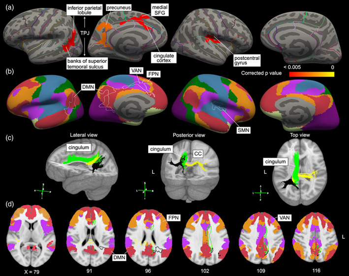
Multiple sclerosis (MS) causes gait and cognitive impairments that are partially normalized by compensatory mechanisms. We aimed to identify the gait tasks that unmask gait disturbance and the underlying neural correlates in MS. We included 25 patients with MS (Expanded Disability Status Scale score: median 2.0, interquartile range 1.0-2.5) and 19 healthy controls. Fast-paced gait examinations with inertial measurement units were conducted, including straight or circular walking with or without cognitive/motor tasks, and the timed up and go test (TUG). Receiver operating characteristic curve analysis was performed to distinguish both groups by the gait parameters. The correlation between gait parameters and cortical thickness or fractional anisotropy values was examined by using three-dimensional T1-weighted imaging and diffusion tensor imaging, respectively (corrected p < .05). Total TUG duration (>6.0 s, sensitivity 88.0%, specificity 84.2%) and stride velocity during cognitive dual-task circular walking (<1.12 m/s, 84.0%, 84.2%) had the highest discriminative power of the two groups. Deterioration of these gait parameters was correlated with thinner cortical thickness in regional areas, including the left precuneus and left temporoparietal junction, overlapped with parts of the default mode network, ventral attention network, and frontoparietal network. Total TUG duration was negatively correlated with fractional anisotropy values in the deep cerebral white matter areas. Turning and multitask gait may be optimal to unveil partially compensated gait disturbance in patients with mild-to-moderate MS through dynamic balance control and multitask processing, based on the structural damage in functional networks.
多发性硬化症 (MS) 可导致步态和认知障碍,部分通过代偿机制得到纠正。我们旨在确定揭示 MS 患者步态障碍及其潜在神经相关性的步态任务。我们纳入了 25 名 MS 患者(扩展残疾状况量表评分中位数为 2.0,四分位距为 1.0-2.5)和 19 名健康对照者。采用惯性测量单元进行快走步态检查,包括直走或圆走,有无认知/运动任务,以及计时起立行走测试(TUG)。通过步态参数对两组进行鉴别,采用受试者工作特征曲线分析。通过三维 T1 加权成像和弥散张量成像分别对步态参数与皮质厚度或各向异性分数值之间的相关性进行检测(校正后 p<0.05)。TUG 总时长(>6.0 s,敏感性 88.0%,特异性 84.2%)和认知双重任务圆走时的步速(<1.12 m/s,84.0%,84.2%)对两组的区分能力最高。这些步态参数的恶化与皮质厚度变薄有关,具体区域包括左侧楔前叶和左侧颞顶联合区,与默认模式网络、腹侧注意网络和额顶叶网络的部分区域重叠。TUG 总时长与深部脑白质区的各向异性分数值呈负相关。基于功能网络的结构损伤,转弯和多任务步态可能是揭示轻度至中度 MS 患者部分代偿性步态障碍的最佳方法,通过动态平衡控制和多任务处理。