Gudas Rimtautas, Staskunas Mantas, Smailys Alfredas, Rimkunas Augustinas
Department of Orthopedics and Traumatology, Hospital of Lithuanian University of Health Sciences (LSMU), Kaunas Clinics, Kaunas, Lithuania.
Department of Orthopedics and Traumatology, Hospital of Lithuanian University of Health Sciences (LSMU), Kaunas Clinics, Kaunas, Lithuania.
Int J Surg Case Rep. 2022 Dec;101:107794. doi: 10.1016/j.ijscr.2022.107794. Epub 2022 Nov 21.
Rare presence of intra-articular osteoid osteoma may be difficult to diagnose due to the lack of typical radiographic features and clinical appearance similar to other articular pathologies. Additionally traditional treatment choices for osteoid osteoma may not suit the given environment of the shoulder joint area.
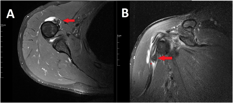
We presented a 50-year-old male with a prolonged history of anterior shoulder pain and shoulder stiffness after physical activity. Intra-articular joint pathology was suspected after initial clinical and radiographic assessment. Magnetic resonance imaging revealed an osteoid osteoma in the humeral bicipital groove.
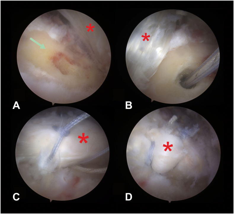
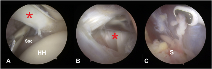
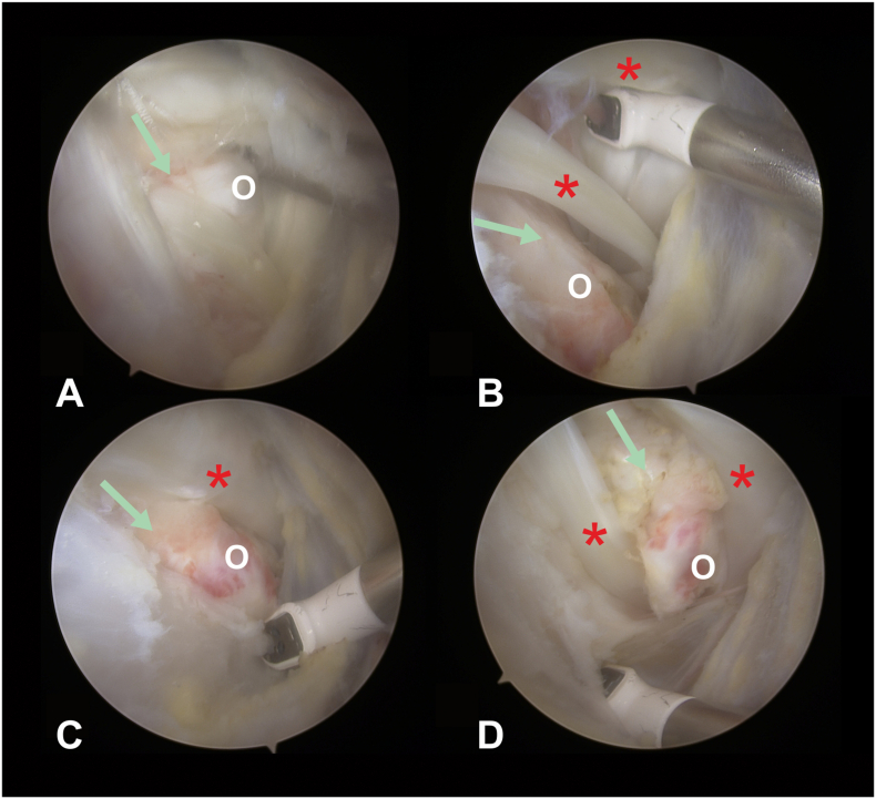
The surgical goal is to resect the benign bony tumour. Though the established treatment by open surgery or radiological minimally invasive techniques may not be optimal since pathologies in the shoulder joint cannot be addressed without the risk of damage to articular structures and increased complications. In this case to avoid joint incision site morbidity and address adjacent pathology arthroscopic removal of the tumour with refixation of the biceps longus tendon was carried out. At follow up of 12 months post-surgery physical activity did not provoke stiffness and resting pain has subsided.
Arthroscopic intra-articular osteoma resection in shoulder joint was optimal to address adjacent osteoma induced pathology, achieve great visualization, reduce incision site complication rates and achieve good results. Additional synovectomy during arthroscopic treatment can be performed, due to concomitant synovitis causing joint stiffness in most reported intra-articular OO cases.
关节内骨样骨瘤罕见,由于缺乏典型的影像学特征且临床表现与其他关节病变相似,可能难以诊断。此外,骨样骨瘤的传统治疗选择可能不适用于肩关节区域的特定情况。
我们报告了一名50岁男性,有长期的前肩部疼痛病史,且在体力活动后出现肩部僵硬。在初步临床和影像学评估后怀疑存在关节内病变。磁共振成像显示肱二头肌沟内有一个骨样骨瘤。
手术目标是切除良性骨肿瘤。尽管通过开放手术或放射学微创技术的既定治疗方法可能并非最佳选择,因为在不损伤关节结构且不增加并发症风险的情况下无法处理肩关节病变。在本病例中,为避免关节切口部位的发病率并处理相邻病变,采用关节镜下切除肿瘤并重新固定肱二头肌长头肌腱。术后12个月随访时,体力活动未引发僵硬,静息痛已消退。
肩关节镜下关节内骨瘤切除术对于处理相邻骨瘤引起的病变、实现良好的可视化、降低切口部位并发症发生率并取得良好效果是最佳选择。由于在大多数报道的关节内骨样骨瘤病例中,伴发的滑膜炎会导致关节僵硬,因此在关节镜治疗期间可进行额外的滑膜切除术。