Department of Histology and Embryology, Akdeniz University School of Medicine, 07070, Antalya, Turkey.
Department of Obstetrics, Gynecology and Reproductive Sciences, Center for Reproductive Sciences, Eli and Edythe Broad Center of Regeneration Medicine and Stem Cell Research, University of California San Francisco, San Francisco, CA, USA.
J Assist Reprod Genet. 2023 Jan;40(1):97-111. doi: 10.1007/s10815-022-02673-z. Epub 2022 Dec 5.
The study aims to investigate first the presence of Syncytin 2 and its receptor, MFSD2, in human sperm, and second whether the expressions of Syncytin 1, Syncytin 2, and their receptors, SLC1A5 and MFSD2, differ between normozoospermic, asthenozoospermic, oligozoospermic, and oligoasthenozoospermic human sperm samples.
The localization patterns and expression levels of syncytins and their receptors were evaluated in normozoospermic (concentration = 88.9 ± 5.5 × 10, motility = 79.2 ± 3.15%, n = 30), asthenozoospermic (concentration = 51.7 ± 7.18 × 10, motility = 24.0 ± 3.12%, n = 15), mild oligozoospermic (concentration = 13.5 ± 2.17 × 10, motility = 72.1 ± 6.5%, n = 15), moderate oligozoospermic (concentration = 8.4 ± 3.21 × 10, motility = 65.1 ± 8.9%, n = 15), severe oligozoospermic (concentration = 2.1 ± 1.01 × 10, motility = 67.5 ± 3.2%, n = 15), and oligoasthenozoospermic (concentration = 5.5 ± 3.21 × 10, motility = 18.5 ± 1.2%, n = 15) samples by immunofluorescence staining and western blot.
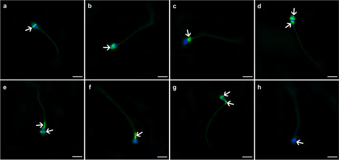
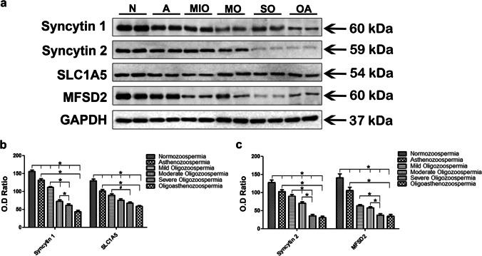
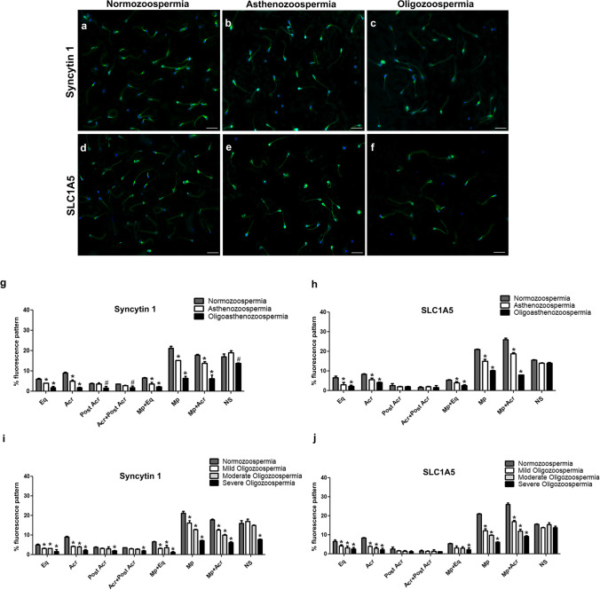
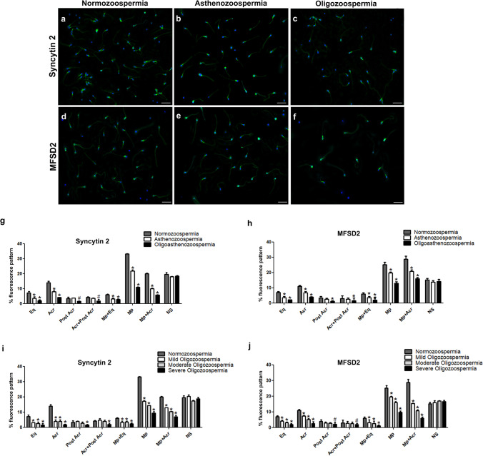
Syncytins and their receptors visualized by immunofluorescence showed similar staining patterns with slight staining of the tail in all spermatozoa regardless of normozoospermia, asthenozoospermia, oligozoospermia, or oligoasthenozoospermia. The localization patterns were categorized as equatorial segment, midpiece region, acrosome, and post-acrosomal areas. The combined staining patterns were also detected as acrosomal cap plus post acrosomal region, the midpiece plus equatorial segment, and midpiece plus acrosomal region. However, some sperm cells were categorized as non-stained. Both syncytin proteins were most intensely localized in the midpiece region, while their receptors were predominantly present in the midpiece plus acrosomal region. Conspicuously, syncytins and their receptors showed decreased expression in asthenozospermic, oligozoospermic, and oligoasthenozoospermic samples compared to normozoospermic samples.
The expression patterns of HERV-derived syncytins and their receptors were identical regardless of the spermatozoa in men with normozoospermia versus impaired semen quality. Further, asthenozoospermia, oligozoospermia, and oligoasthenozoospermia as male fertility issues are associated with decreased expression of both syncytins and their receptors.
本研究旨在首先探讨 Syncytin 2 及其受体 MFSD2 是否存在于人类精子中,其次研究 Syncytin 1、Syncytin 2 及其受体 SLC1A5 和 MFSD2 的表达是否存在差异在正常精子、弱精子、少精子和少弱精子的人类精子样本中。
通过免疫荧光染色和 Western blot 分析,评估同步蛋白及其受体在正常精子(浓度=88.9±5.5×10 个/ml,活力=79.2±3.15%,n=30)、弱精子(浓度=51.7±7.18×10 个/ml,活力=24.0±3.12%,n=15)、轻度少精子(浓度=13.5±2.17×10 个/ml,活力=72.1±6.5%,n=15)、中度少精子(浓度=8.4±3.21×10 个/ml,活力=65.1±8.9%,n=15)、严重少精子(浓度=2.1±1.01×10 个/ml,活力=67.5±3.2%,n=15)和少弱精子(浓度=5.5±3.21×10 个/ml,活力=18.5±1.2%,n=15)样本中的定位模式和表达水平。
免疫荧光染色显示,同步蛋白及其受体的可视化显示出相似的染色模式,所有精子尾部都有轻微染色,无论是否为正常精子、弱精子、少精子还是少弱精子。定位模式分为赤道段、中段、顶体和顶体后区。联合染色模式也被检测为顶体帽加顶体后区、中段加赤道段和中段加顶体区。然而,一些精子细胞被归类为未染色。两种 Syncytin 蛋白主要定位于中段区域,而它们的受体主要存在于中段加顶体区域。值得注意的是,与正常精子样本相比,弱精子症、少精子症和少弱精子症样本中的 Syncytin 蛋白及其受体表达明显降低。
在具有正常精子和受损精液质量的男性中,HERV 衍生的 Syncytin 及其受体的表达模式是相同的。此外,弱精子症、少精子症和少弱精子症等男性生育问题与 Syncytin 及其受体的表达降低有关。