Orthopaedic Clinic CTO, University of Florence, Largo Palagi 1, 50139, Florence, Italy.
J Med Case Rep. 2022 Dec 10;16(1):457. doi: 10.1186/s13256-022-03667-2.
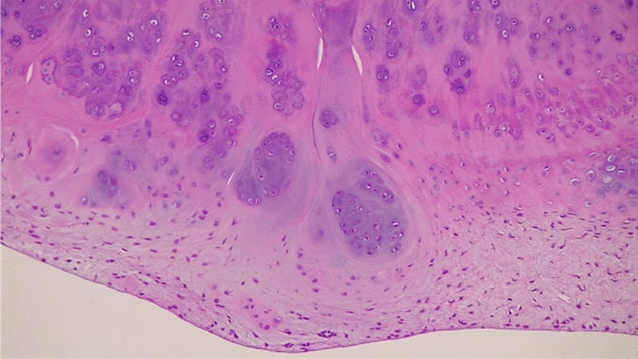
The synovial chondromatosis is an uncommon proliferative metaplastic process of the synovial cells that can develop in any synovial joint. An isolated primary chondromatosis of the posterior compartment of the knee is uncommon and few cases are reported in literature. Our purpose is to describe a rare case of primary chondromatosis of the knee posterior compartment and report the arthroscopic loose bodies excision through a difficult posteromedial portal, avoiding the use of the accessory posterior portal, most commonly reported for approaching this disease.
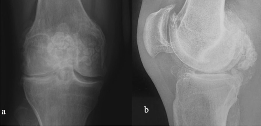
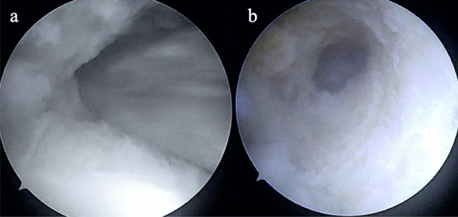
We report a rare case of a 35-year-old Caucasian male patient with diagnosis of chondromatosis of the posterior knee compartment. The radiographs showed multiple loose bodies of the posterior compartment. The MRI revealed minimal synovial hypertrophy areas, multiple osteophytes in the intercondylar notch, and loose bodies in the posteromedial compartment. The CT allowed us to assess the bony structures, the morphology of the intercondylar notch, and the presence osteophytes of the medial and lateral femoral condyles. The CT images were crucial to plan how to reach the posterior compartments of the knee through a trans-notch passage. The patient underwent arthroscopic surgery using anteromedial, anterolateral, and posteromedial portals. The tunneling through the intercondylar osteophytes was performed to allow the arthroscope to pass trans-notch. To avoid additional accessory posterior portals, we used a 70° arthroscope to better explore the posterior knee compartment. The cartilage-like bodies were removed and synovectomy of the inflamed areas was performed. The clinical and radiological follow-up was 12 months and the patient showed excellent clinical outcomes, returning to his activities of daily living and sport activity.
Our case report highlights the importance of the arthroscopic approach to treat synovial chondromatosis, despite the involvement of the posterior compartment of the knee. An optimal preoperative imaging allows to plan for the proper surgical procedure even in patients with severe osteoarthritis. Moreover, the adoption of an intercondylar notch tunneling and a 70° arthroscope can help surgeons to better explore the posterior knee compartment, avoiding an accessory posterior trans-septal portal. Therefore, a synovectomy of the inflamed foci may be useful to prevent recurrence.
滑膜软骨瘤病是一种少见的滑膜细胞增生性化生过程,可发生于任何滑膜关节。孤立性膝关节后关节室原发性软骨瘤病少见,文献报道病例较少。我们的目的是描述一例罕见的膝关节后关节室原发性软骨瘤病,并报告通过一个困难的后内侧入路关节镜下切除游离体,避免使用最常报道的辅助后关节室间入路来治疗这种疾病。
我们报告了一例罕见的 35 岁白人男性患者,诊断为膝关节后关节室软骨瘤病。X 线片显示后关节室多个游离体。MRI 显示轻微的滑膜增生区、髁间窝内多个骨赘和后内侧关节室的游离体。CT 有助于评估骨结构、髁间窝形态和内外侧股骨髁骨赘的存在。CT 图像对于规划如何通过关节间隙入路到达膝关节后关节室至关重要。患者接受了关节镜手术,采用前内侧、前外侧和后内侧入路。通过骨赘隧道化使关节镜通过关节间隙。为避免额外的辅助后关节室间入路,我们使用 70°关节镜更好地探查膝关节后关节室。切除软骨样体并对炎症区域进行滑膜切除术。临床和放射学随访 12 个月,患者临床结果良好,恢复日常生活和运动活动。
尽管涉及膝关节后关节室,我们的病例报告强调了关节镜治疗滑膜软骨瘤病的重要性。术前的最佳影像学检查有助于规划适当的手术程序,即使是在严重骨关节炎的患者中。此外,采用髁间窝隧道化和 70°关节镜可以帮助外科医生更好地探查膝关节后关节室,避免使用辅助后关节室间隔入路。因此,滑膜切除术可能有助于预防复发。