Aili Yirizhati, Maimaitiming Nuersimanguli, Maimaiti Aierpati, Liu Wen, Qin Hu, Ji Wenyu, Mahemuti Yusufu, Wang Yongxin, Wang Zengliang
Department of Neurosurgery, The First Affiliated Hospital of Xinjiang Medical University, Urumqi, Xinjiang, China.
Department of Oncology, The First Affiliated Hospital of Xinjiang Medical University, Urumqi, Xinjiang, China.
J Oncol. 2022 Nov 30;2022:2621969. doi: 10.1155/2022/2621969. eCollection 2022.
VASH1 is a novel angiogenic regulatory factor, that participates in the process of carcinogenesis and the development of diverse tumors. Our study aimed to investigate the expression and prognostic value of the VASH1 in Lower-Grade Glioma (LGG), to explore its functional network in LGG and its effects on biological behaviors.
LGG transcriptome data, somatic mutation profiles and clinical features analyzed in the present study were obtained from the TCGA, GTEx, CCLE, CGGA, UALCAN, and GEPIA2 databases, as well as clinical data and tissue sections of 83 LGG patients in our hospital. The expression characteristics of VASH1 in LGG were investigated by univariate, multivariate, immunohistochemistry, qRT-PCR, and western-blot. Subsequently, we analyzed the prognostic significance of VASH1 in LGG patients by survival analysis, subject operation characteristic curve, correlation analysis, external validation, independent prognostic significance analysis, and clinical stratification, and confirmed its biological effect on glioma cell lines in vitro. Finally, we performed GO, KEGG, and GSEA to clarify biological functions and related pathways. CIBERSORT and ESTIMATE algorithms were used to calculate the proportion of immune cells and immune microenvironment fraction in LGG.
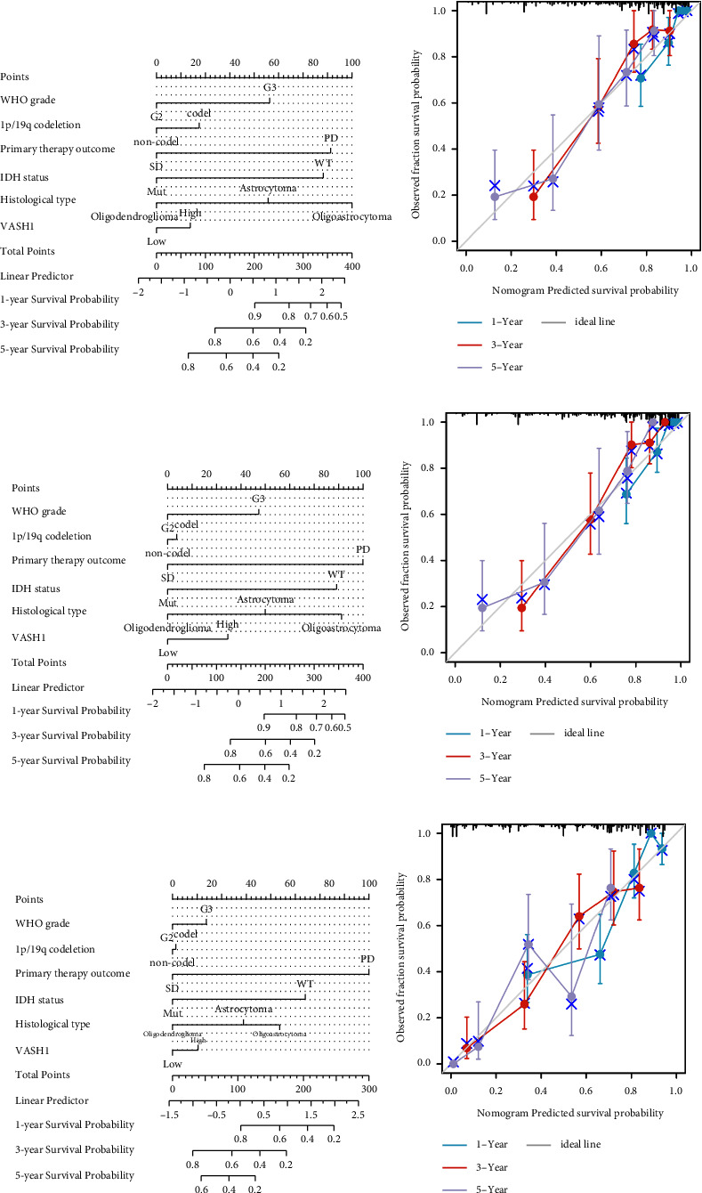
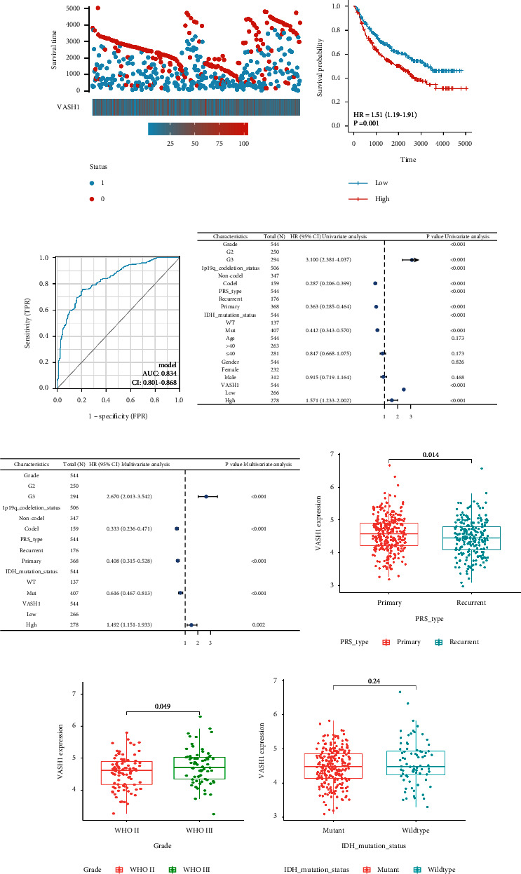
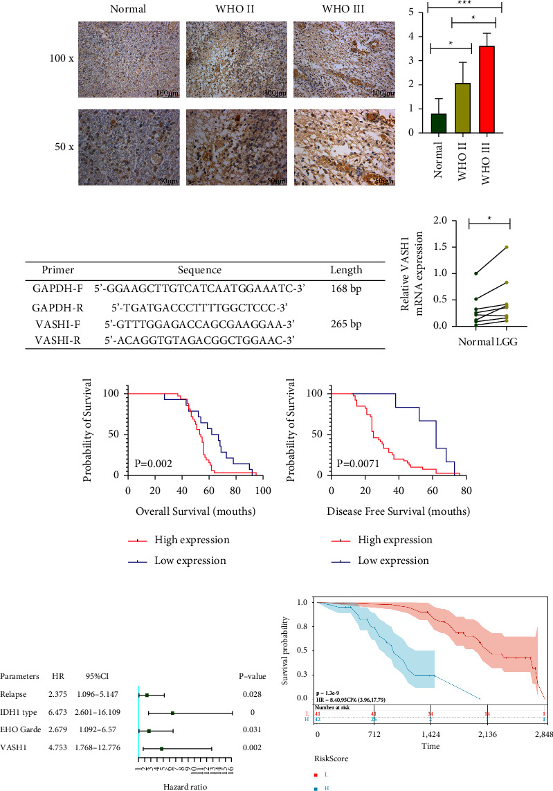
We found that VASH1 is highly expressed in LGG tissues and is associated with poor prognosis, WHO grade, IDH1 wild-type, and progressive disease ( < 0.05). Multivariate and the Nomogram model showed that high VASH1 expression was an independent risk factor for glioma prognosis and had better prognostic prediction efficacy in different LGG Patient cohorts (HR = 4.753 and =0.002). In vitro experiments showed that knockdown of VASH1 expression in glioma cell lines caused increased glioma cell proliferation, invasion, and migration capacity. The mechanism may be related to VASH1 promoting microtubule formation and remodeling of immune microenvironment.
Our study firstly found that high VASH1 expression was associated with poor prognosis. In addition, We identified the possible mechanism by which VASH1 functioned in LGG. VASH1 inhibits the invasion and migration of tumor cells by affecting microtubule formation and immune infiltration in the tumor microenvironment. May be an important endogenous anti-tumor factor for LGG and provide a potential biomarker for individualized treatment of LGG.
VASH1是一种新型血管生成调节因子,参与肿瘤发生过程及多种肿瘤的发展。本研究旨在探讨VASH1在低级别胶质瘤(LGG)中的表达及预后价值,探索其在LGG中的功能网络及其对生物学行为的影响。
本研究分析的LGG转录组数据、体细胞突变谱和临床特征来自TCGA、GTEx、CCLE、CGGA、UALCAN和GEPIA2数据库,以及我院83例LGG患者的临床资料和组织切片。通过单因素、多因素、免疫组织化学、qRT-PCR和western-blot研究VASH1在LGG中的表达特征。随后,我们通过生存分析、受试者操作特征曲线、相关性分析、外部验证、独立预后意义分析和临床分层分析VASH1在LGG患者中的预后意义,并在体外证实其对胶质瘤细胞系的生物学作用。最后,我们进行GO、KEGG和GSEA以阐明生物学功能和相关途径。使用CIBERSORT和ESTIMATE算法计算LGG中免疫细胞比例和免疫微环境分数。
我们发现VASH1在LGG组织中高表达,且与预后不良、WHO分级、IDH1野生型和疾病进展相关(<0.05)。多因素和列线图模型显示,VASH1高表达是胶质瘤预后的独立危险因素,在不同LGG患者队列中具有更好的预后预测效能(HR = 4.753,=0.002)。体外实验表明,敲低胶质瘤细胞系中VASH1的表达会导致胶质瘤细胞增殖、侵袭和迁移能力增强。其机制可能与VASH1促进微管形成和免疫微环境重塑有关。
我们的研究首次发现VASH1高表达与预后不良相关。此外,我们确定了VASH1在LGG中发挥作用的可能机制。VASH1通过影响微管形成和肿瘤微环境中的免疫浸润来抑制肿瘤细胞的侵袭和迁移。可能是LGG重要的内源性抗肿瘤因子,并为LGG的个体化治疗提供潜在生物标志物。