Clinical Neurosciences, Department of Clinical Medicine, University of Turku, Turku, Finland.
Neurocenter, Turku University Hospital, Turku, Finland.
BMC Neurol. 2022 Dec 15;22(1):487. doi: 10.1186/s12883-022-03007-3.
Leber's hereditary optic neuropathy (LHON) is a common form of mitochondrial disease. The typical clinical presentation of LHON is subacute, painless loss of vision resulting from bilateral optic nerve atrophy. Moreover, extra-ocular manifestations such as cardiac conduction abnormalities and neurological manifestations such as multiple sclerosis (MS) like disease or parkinsonism are encountered in some patients. Abnormal findings in spinal cord MR imaging or in the cerebrospinal fluid (CSF) have been observed in previous cases of LHON-associated myelopathy.
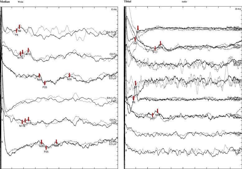
We report a male patient with LHON who developed symptoms of myelopathy including gait unsteadiness, enhanced deep tendon reflexes and sensory loss of the lower extremities. Imaging of the brain and spinal cord, CSF analysis, as well as neurography and electromyography did not disclose any abnormalities. The somatosensory evoked potential (SEP) findings were suggestive of dorsal column dysfunction.
The patient case demonstrates that myelopathy associated with LHON can present without abnormal findings in central nervous system MR imaging or in the CSF, and without evidence suggestive of multiple sclerosis or MS-like disease. The dorsal column seems to be particularly vulnerable to myelopathy changes in LHON. Evoked potential investigations may assist in confirming the diagnosis, when clinical features are in line with myelopathy but findings in CSF analysis and central nervous system imaging are normal.
Leber 遗传性视神经病变(LHON)是一种常见的线粒体疾病。LHON 的典型临床表现为亚急性、无痛性视力丧失,由双侧视神经萎缩引起。此外,一些患者还会出现心脏传导异常等眼外表现和多发性硬化症(MS)样疾病或帕金森病等神经系统表现。在以前的 LHON 相关脊髓病病例中,已经观察到脊髓磁共振成像或脑脊液(CSF)中的异常发现。
我们报告了一例男性 LHON 患者,出现了包括步态不稳、深腱反射增强和下肢感觉丧失在内的脊髓病症状。脑和脊髓成像、CSF 分析以及神经图和肌电图均未发现任何异常。体感诱发电位(SEP)检查结果提示背柱功能障碍。
该患者病例表明,与 LHON 相关的脊髓病可在中枢神经系统磁共振成像或 CSF 中无异常发现,并且无多发性硬化症或 MS 样疾病的证据。在 LHON 中,背柱似乎特别容易发生脊髓病变化。当临床特征符合脊髓病,但 CSF 分析和中枢神经系统成像结果正常时,诱发电位检查可能有助于确认诊断。