Department of Radiology, The Affiliated Hospital of Qingdao University, Qingdao, China.
Department of Stomatology, The Affiliated Hospital of Qingdao University, Qingdao, China.
BMC Neurol. 2022 Dec 22;22(1):498. doi: 10.1186/s12883-022-03026-0.
The MRI features of Diffuse midline glioma, H3 K27-altered and glioma in the midline without H3 K27-altered were compared and analyzed, and the changes in the apparent diffusion coefficient (ADC) of the two groups were quantitatively analyzed.
The MRI images of 35 patients with Diffuse midline gliomas, H3 K27-altered and gliomas in the midline without H3 K27-altered were analyzed retrospectively. The location, edge, signal, peritumoral edema and enhancement characteristics of the lesions were observed, and the changes in ADC values were analyzed.
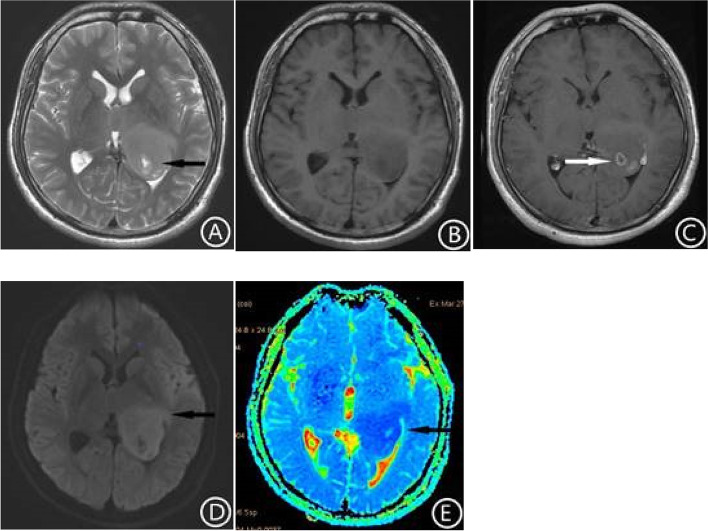
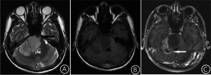
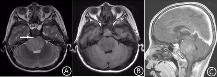
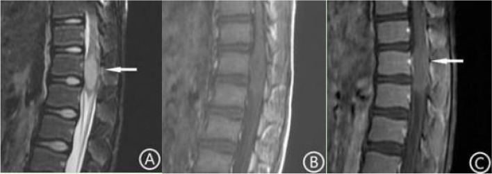
In the H3 K27-altered group, 85.7% (12/14) of the tumors were located in the thalamus and brainstem compared with 28.6% (6/21) in the no H3 K27-altered group. In the H3 K27-altered group, for tumors only located in the midline area, only 14.3% (1/7) had irregular shapes and unclear boundaries, while for tumors also invaded the extramidline tissues 85.7% (6/7) had irregular shapes and unclear boundaries.The"basilar artery wrapped sign" was found in 6 patients with tumors located in the pons in the H3 K27-altered group, but none in the no H3 K27-altered group had this sign. In the H3 K27-altered group, only 14.3% (1/7) of the tumors confined to the midline area had small cystic degeneration and necrosis, while for tumors also invaded the extramidline tissues, 100% (7/7) of the tumors had cystic degeneration and necrosis, and the cystic degeneration and necrosis only located in the extramidline region of the tumor in 6 cases.A total of 78.6% (11/14) of tumors in the H3 K27-altered group showed mild to moderate enhancement, while 47.6% (10/21) of tumors in the no H3 K27-altered group showed mild to moderate enhancement. The average peritumoral edema index was 1.13 in the H3 K27-altered group and 1.75 in the no H3 K27-altered group. The average ADC value of tumor in the H3 K27-altered group was 7.83 × 10 mm/s, and the ratio to normal brain tissue was 0.844, while the values in the no H3 K27-altered group were 13.5 × 10 mm/s and 1.75, respectively.
Compared with gliomas in the midline without H3 K27-altered, The MRI findings and ADC value of Diffuse midline gliomas, H3K27-altered have some characteristics, which can help improve the diagnosis and differential diagnosis.
比较并分析弥漫性中线胶质瘤 H3 K27 改变型和中线无 H3 K27 改变型胶质瘤的 MRI 特征,并对两组表观扩散系数(ADC)的变化进行定量分析。
回顾性分析 35 例弥漫性中线胶质瘤 H3 K27 改变型和中线无 H3 K27 改变型胶质瘤患者的 MRI 图像。观察病变的位置、边缘、信号、瘤周水肿和强化特征,并分析 ADC 值的变化。
在 H3 K27 改变型组中,85.7%(12/14)的肿瘤位于丘脑和脑干,而在无 H3 K27 改变型组中仅为 28.6%(6/21)。在 H3 K27 改变型组中,仅中线区域的肿瘤,形态不规则和边界不清的比例为 14.3%(1/7),而同时侵犯中线外组织的肿瘤,形态不规则和边界不清的比例为 85.7%(6/7)。在 H3 K27 改变型组中,有 6 例桥脑肿瘤存在“基底动脉包绕征”,而无 H3 K27 改变型组均无此征象。在 H3 K27 改变型组中,仅中线区域肿瘤局限的肿瘤小囊变坏死比例为 14.3%(1/7),而同时侵犯中线外组织的肿瘤,囊变坏死比例为 100%(7/7),其中 6 例肿瘤的囊变坏死仅位于肿瘤中线外区域。H3 K27 改变型组肿瘤的强化程度为轻度至中度的比例为 78.6%(11/14),无 H3 K27 改变型组肿瘤的强化程度为轻度至中度的比例为 47.6%(10/21)。H3 K27 改变型组的平均瘤周水肿指数为 1.13,无 H3 K27 改变型组为 1.75。H3 K27 改变型组肿瘤的平均 ADC 值为 7.83×10mm/s,与正常脑组织的比值为 0.844,而无 H3 K27 改变型组肿瘤的平均 ADC 值为 13.5×10mm/s,与正常脑组织的比值为 1.75。
与中线无 H3 K27 改变型胶质瘤相比,弥漫性中线胶质瘤 H3 K27 改变型的 MRI 表现和 ADC 值具有一定特征,有助于提高诊断和鉴别诊断水平。