Matveenko Andrew G, Ryzhkova Varvara E, Zaytseva Natalia A, Danilov Lavrentii G, Mikhailichenko Anastasia S, Barbitoff Yury A, Zhouravleva Galina A
Department of Genetics and Biotechnology, St. Petersburg State University, 199034 St. Petersburg, Russia.
Laboratory of Amyloid Biology, St. Petersburg State University, 199034 St. Petersburg, Russia.
Biology (Basel). 2022 Nov 22;11(12):1688. doi: 10.3390/biology11121688.
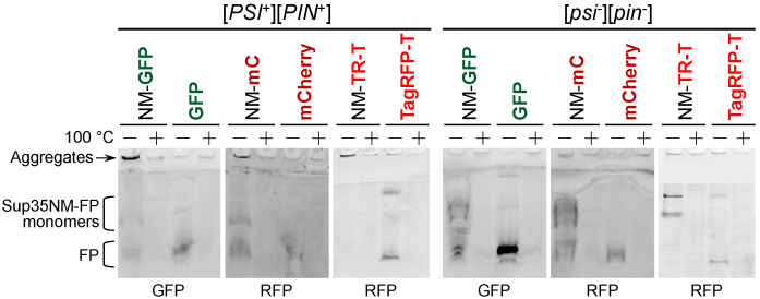
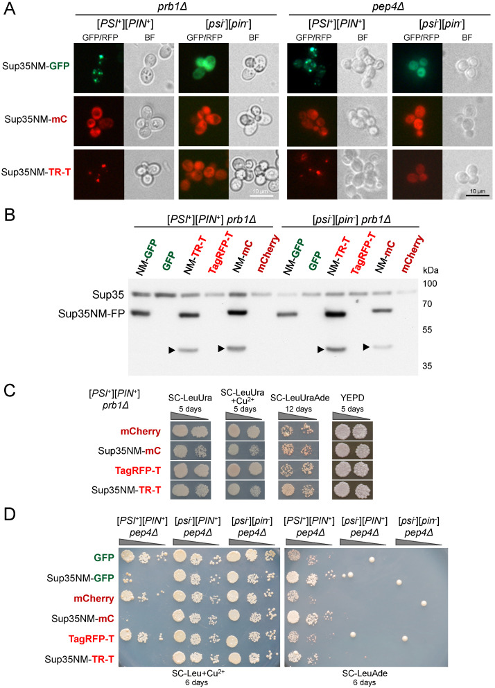
Yeast is a convenient model for studying protein aggregation as it is known to propagate amyloid prions. [] is the prion form of the release factor eRF3 (Sup35). Aggregated Sup35 causes defects in termination of translation, which results in nonsense suppression in strains carrying premature stop codons. N-terminal and middle (M) domains of Sup35 are necessary and sufficient for maintaining [] in cells while preserving the prion strain's properties. For this reason, Sup35NM fused to fluorescent proteins is often used for [] detection and investigation. However, we found that in such chimeric constructs, not all fluorescent proteins allow the reliable detection of Sup35 aggregates. Particularly, transient overproduction of Sup35NM-mCherry resulted in a diffuse fluorescent pattern in the [] cells, while no loss of prions and no effect on the Sup35NM prion properties could be observed. This effect was reproduced in various unrelated strain backgrounds and prion variants. In contrast, Sup35NM fused to another red fluorescent protein, TagRFP-T, allowed the detection of [] aggregates. Analysis of protein lysates showed that Sup35NM-mCherry is actively degraded in the cell. This degradation was not caused by vacuolar proteases and the ubiquitin-proteasomal system implicated in the Sup35 processing. Even though the intensity of this proteolysis was higher than that of Sup35NM-GFP, it was roughly the same as in the case of Sup35NM-TagRFP-T. Thus, it is possible that, in contrast to TagRFP-T, degradation products of Sup35NM-mCherry still preserve their fluorescent properties while losing the ability to decorate pre-existing Sup35 aggregates. This results in diffuse fluorescence despite the presence of the prion aggregates in the cell. Thus, tagging with fluorescent proteins should be used with caution, as such proteolysis may increase the rate of false-negative results when detecting prion-bearing cells.
酵母是研究蛋白质聚集的便捷模型,因为已知它能传播淀粉样朊病毒。[]是释放因子eRF3(Sup35)的朊病毒形式。聚集的Sup35会导致翻译终止缺陷,这会在携带提前终止密码子的菌株中导致无义抑制。Sup35的N端和中间(M)结构域对于在细胞中维持[]并保持朊病毒株的特性是必要且充分的。因此,与荧光蛋白融合的Sup35NM常用于[]检测和研究。然而,我们发现,在这种嵌合构建体中,并非所有荧光蛋白都能可靠地检测到Sup35聚集体。特别是,Sup35NM-mCherry的瞬时过量表达在[]细胞中导致弥散的荧光模式,而未观察到朊病毒丢失且对Sup35NM朊病毒特性无影响。这种效应在各种不相关的菌株背景和朊病毒变体中都能重现。相比之下,与另一种红色荧光蛋白TagRFP-T融合的Sup35NM能够检测到[]聚集体。蛋白质裂解物分析表明,Sup35NM-mCherry在细胞中被积极降解。这种降解不是由液泡蛋白酶和参与Sup35加工的泛素-蛋白酶体系统引起的。尽管这种蛋白水解的强度高于Sup35NM-GFP,但与Sup35NM-TagRFP-T的情况大致相同。因此,与TagRFP-T不同,Sup35NM-mCherry的降解产物可能仍保留其荧光特性,同时失去装饰预先存在的Sup35聚集体的能力。这导致尽管细胞中存在朊病毒聚集体,但仍出现弥散荧光。因此,用荧光蛋白进行标记时应谨慎,因为这种蛋白水解可能会增加检测含朊病毒细胞时假阴性结果的发生率。