Department of Clinical Medicine, UiT The Arctic University, Tromso, Norway.
Department of Cardiology, Division of Cardiothoracic and Respiratory Medicine, University Hospital of North Norway, Tromso, Norway.
Open Heart. 2022 Dec;9(2). doi: 10.1136/openhrt-2022-002136.
Strain artefacts are known to hamper the correct interpretation of segmental strain and strain-rate (S/SR). Defining the normal ranges of myocardial segmental deformation is important in clinical studies and routine echocardiographic practice. In order to define artefact-free normal ranges for segmental longitudinal S/SR parameters, we investigated the extent to which different types of artefacts and their segmental localisation in the three different myocardial layers created a bias in the results of echocardiographic strain measurements.
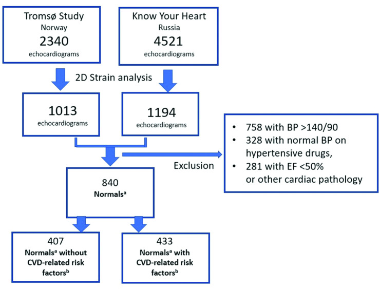
The study included echocardiograms from men and women aged 40-69 years from two population-based studies, namely the Know Your Heart study (Russia) and the Tromsø Study (Norway). Of the 2207 individuals from these studies, 840 had normal results, defined as the absence of hypertension or indicators of any cardiovascular disease. Two-dimensional (2D) global and segmental S/SR of the three myocardial layers were analysed using speckle tracking echocardiography. Artefacts were assessed with two different methods: visual identification of image-artefacts and a novel conceptual approach of 'curve-artefacts' or unphysiological strain-curve formation.
Segmental strain values were found to have significantly reduced in the presence of strain-curve artefacts (14.9%±5.8% towards -20.7%±4.9%), and increased with the foreshortening of the 2D image. However, the individual global strain values were not substantially altered by discarding segmental artefacts. Reduction due to artefacts was observed in all segments, layers, systolic and diastolic strain, and SR. Thus, we presented normal ranges for basal-septal, basal, medial and apical segment groups after excluding artefacts.
Strain-curve artefacts introduce systematic errors, resulting in reduced segmental S/SR values. In terms of artefact-robust global longitudinal strain, the detection of curve-artefacts is crucial for the correct interpretation of segmental S/SR patterns. Intersegmental S/SR gradients and artefacts need to be considered for the correct definition of normalcy and pathology.
众所周知,应变伪影会影响节段应变和应变速率(S/SR)的正确解读。定义心肌节段变形的正常范围对于临床研究和常规超声心动图实践非常重要。为了定义节段纵向 S/SR 参数无伪影的正常范围,我们研究了不同类型的伪影及其在三个不同心肌层中的节段定位对超声心动图应变测量结果产生的偏差程度。
该研究纳入了来自俄罗斯的“了解你的心脏”研究(Know Your Heart study)和挪威的特罗姆瑟研究(Tromsø Study)这两项基于人群的研究中年龄在 40-69 岁的男性和女性的超声心动图。在这两项研究的 2207 名个体中,840 名个体的结果正常,定义为无高血压或任何心血管疾病的指标。使用斑点追踪超声心动图分析三个心肌层的二维(2D)整体和节段 S/SR。使用两种不同的方法评估伪影:图像伪影的视觉识别和新颖的“曲线伪影”或非生理应变曲线形成的概念方法。
存在应变曲线伪影时,节段应变值显著降低(从 14.9%±5.8%降至-20.7%±4.9%),并且随着 2D 图像的缩短而增加。然而,通过丢弃节段伪影,个体整体应变值并没有实质性改变。所有节段、层、收缩期和舒张期应变以及 SR 都观察到由于伪影导致的降低。因此,在排除伪影后,我们为基底隔段、基底段、中层和心尖段组提供了正常范围。
应变曲线伪影会引入系统误差,导致节段 S/SR 值降低。就具有抗伪影能力的整体纵向应变而言,检测曲线伪影对于正确解读节段 S/SR 模式至关重要。为了正确定义正常和病理,需要考虑节段间 S/SR 梯度和伪影。