The Department of Urology, Affiliated Sixth People's Hospital, Shanghai Jiaotong University School of Medicine, No. 600, Yishan Road, Shanghai, China.
School of Materials Science and Engineering, Shanghai University of Engineering Science, Shanghai, China.
BMC Urol. 2023 Jan 6;23(1):1. doi: 10.1186/s12894-022-01165-7.
Urethral stenosis caused by pelvic fracture urethral injury (PFUI) is a complex urological disease, especially for the redo cased. However, to find the proximal end of the posterior urethra, and to avoid injury to the rectum and to forecast to remove the inferior pubic margin are two key points for a successful surgery. These steps can be challenging for even the most experienced urologists. This study is to describe a new technique for understanding the three-dimensional (3D) anatomy of the urethra, which will also aid in surgical planning and simplify urethroplasty.
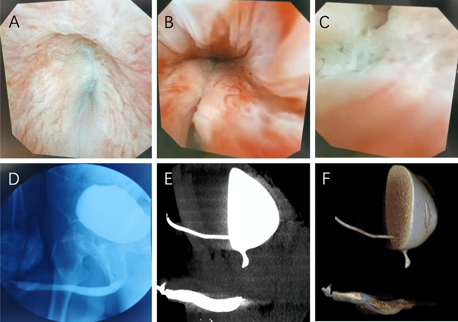
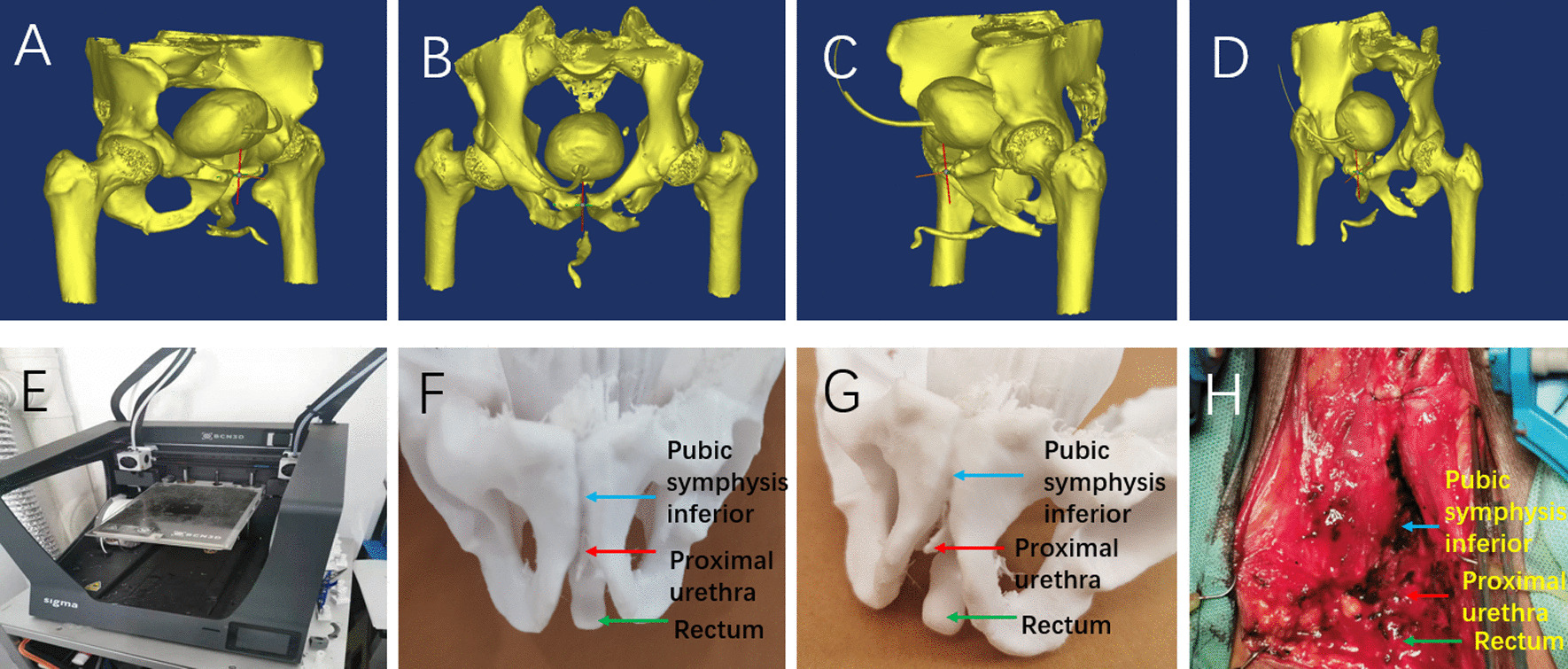
Three patients underwent routine urethroscopy, X ray urethrography and contrast CT urethrography. The 3D images were then reconstructed, and the data were transmitted to a 3D printer. 3D models were printed with polyacrylic acid to simulate the anatomical structure and relationship of urethral stenosis with pubic symphysis and rectum. Various diagnosis methods were compared with the condition in surgery. The patients and trainee questionnaires were performed.
Three models of urethral CT were obtained. These models were presented to patients and trainee doctors along with routine urethroscopy, urethrography, and urethral CT. The scores of patients and trainee question forms demonstrated that the 3D printed urethral stenosis model of pelvic fracture has obvious advantages in urethral adjacency and ease of understanding. The 3D printed urethras were easy to show the pubic symphysis and simulate its excision and exposure of urethra. The model could show the precise distance from urethra to rectum to prevent the rectum injury in surgery.
3D printing technology can be applied to the preoperative evaluation of urethral stenosis caused by PFUI. It can be auxiliary to understand the anatomical structure of the posterior urethra, the direction of urethral displacement, protecting the rectum and the forecasting for pubectomy. It is especially helpful for the accurate preoperative planning of some complex urethral stenosis and redo cases.
骨盆骨折尿道损伤(PFUI)导致的尿道狭窄是一种复杂的泌尿科疾病,尤其是对于再发性病例。然而,找到后尿道的近端,并避免损伤直肠,预测并切除耻骨下支,这两点是手术成功的关键。即使是最有经验的泌尿科医生,这些步骤也具有挑战性。本研究旨在描述一种新的技术,以了解尿道的三维(3D)解剖结构,这也将有助于手术规划并简化尿道成形术。
对 3 例患者进行常规尿道镜检查、X 射线尿道造影和对比 CT 尿道造影。然后重建 3D 图像,并将数据传输到 3D 打印机。使用聚丙烯酸打印 3D 模型,以模拟尿道狭窄与耻骨联合和直肠的解剖结构和关系。将各种诊断方法与手术中的情况进行比较。对患者和受训者进行问卷调查。
获得了 3 例尿道 CT 模型。这些模型与常规尿道镜检查、尿道造影和尿道 CT 一起呈现给患者和受训医生。患者和受训者问卷调查表的评分表明,骨盆骨折 3D 打印尿道狭窄模型在尿道毗邻和易于理解方面具有明显优势。3D 打印尿道易于显示耻骨联合,并模拟其切除和暴露尿道。该模型可以显示尿道与直肠的精确距离,以防止手术中直肠损伤。
3D 打印技术可应用于 PFUI 所致尿道狭窄的术前评估。它可以辅助了解后尿道的解剖结构、尿道移位的方向、保护直肠和预测耻骨切除。对于一些复杂的尿道狭窄和再发性病例,它尤其有助于进行准确的术前规划。