Abdominal Oncology Ward, Cancer Center, West China Hospital, Sichuan University, Chengdu, China.
Cancer Center, Sichuan Provincial People's Hospital, University of Electronic Science and Technology of China (Chinese Academy of Sciences Sichuan Translational Medicine Research Hospital), Chengdu, China.
Dig Dis Sci. 2023 Jul;68(7):3070-3082. doi: 10.1007/s10620-023-07824-5. Epub 2023 Jan 21.
Ferroptosis, as a unique form of cell death, plays crucial negative roles in tumorigenesis and progression. This study aimed to investigate the role and molecular mechanism of TEA domain transcription factor 1 (TEAD1) in HCC and its effect on sorafenib-induced ferroptosis.
TEAD1 expression was analyzed in HCC tissues using quantitative PCR, and western blot. The effects on cell proliferation, migration and invasion were determined by CCK-8, wound healing and Transwell assays. Intracellular iron, reactive oxygen species (ROS), malondialdehyde (MDA) and GSH measurement was used to assess ferroptosis. Chromatin immunoprecipitation and luciferase reporter gene assays were performed to verify the relationship between TEAD1 and solute carrier family 3 member 2 (SLC3A2). Expression of mTOR, ribosomal protein S6, glutathione peroxidase 4 (GPX4) and SLC3A2 was analyzed by western blot. Tumor xenografts were used assess the effect of TEAD1 on tumor growth in vivo.
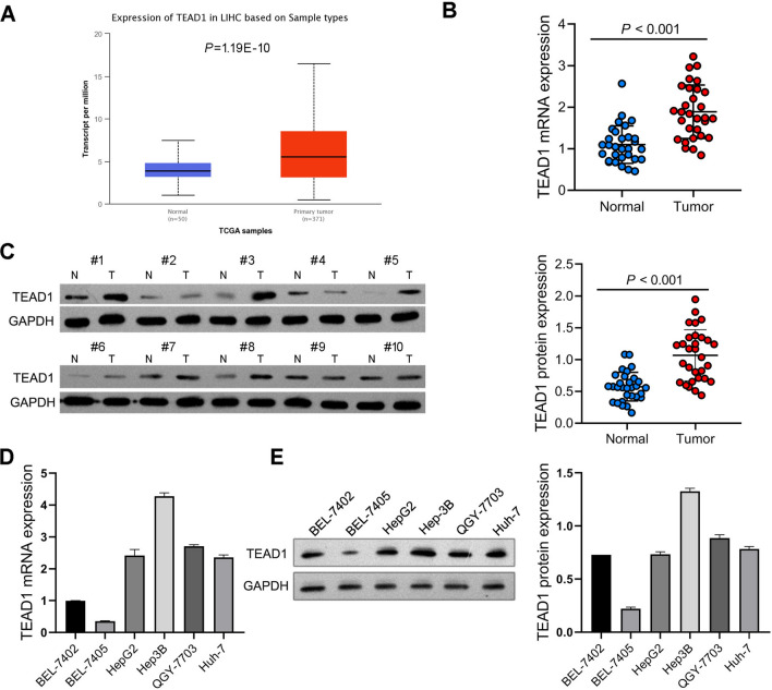
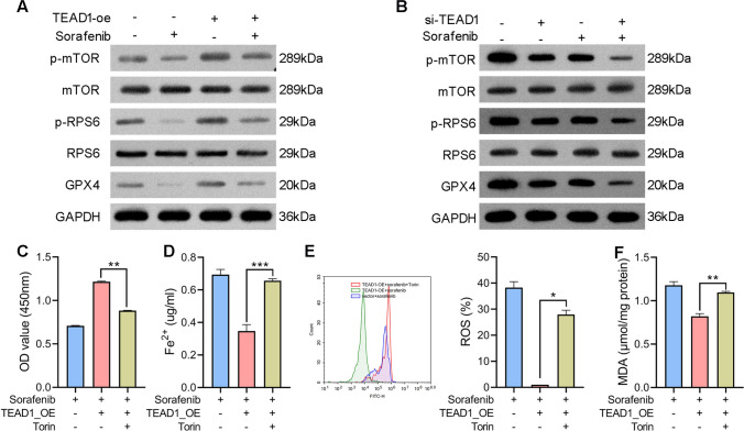
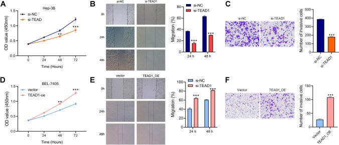
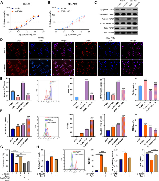
TEAD1 was more abundant in HCC compared with normal tissues. Overexpression of TEAD1 enhanced the proliferation, migration, and invasion of HCC cells, while knockdown of TEAD1 inhibited these cell behaviors. Further, TEAD1 inhibited ferroptosis, which was demonstrated by decreased intracellular Fe content, ROS, and MDA levels, and increased GSH activity. Mechnistically, TEAD1 promotes the transcription of SLC3A2 and activates the mTOR signaling. Additionally, silenced TEAD1 restrained tumor growth and enhance sorafenib-induced antitumor activity in vivo.
TEAD1 confers resistance of HCC cells to ferroptosis, thereby promoting the progression of HCC, suggesting the potential value of TEAD1 in the diagnosis and treatment of HCC.
铁死亡作为一种独特的细胞死亡形式,在肿瘤发生和进展中起着关键的负向作用。本研究旨在探讨 TEA 结构域转录因子 1(TEAD1)在肝癌中的作用及其对索拉非尼诱导铁死亡的影响及其分子机制。
采用实时定量 PCR 和 Western blot 分析肝癌组织中 TEAD1 的表达。通过 CCK-8 法、划痕愈合实验和 Transwell 实验检测细胞增殖、迁移和侵袭的影响。通过细胞内铁、活性氧(ROS)、丙二醛(MDA)和 GSH 测定评估铁死亡。通过染色质免疫沉淀和荧光素酶报告基因检测验证 TEAD1 与溶质载体家族 3 成员 2(SLC3A2)的关系。Western blot 分析 mTOR、核糖体蛋白 S6、谷胱甘肽过氧化物酶 4(GPX4)和 SLC3A2 的表达。采用肿瘤异种移植评估 TEAD1 对体内肿瘤生长的影响。
TEAD1 在肝癌组织中的表达高于正常组织。过表达 TEAD1 增强了肝癌细胞的增殖、迁移和侵袭能力,而 TEAD1 敲低则抑制了这些细胞行为。此外,TEAD1 抑制了铁死亡,表现为细胞内铁含量、ROS 和 MDA 水平降低,GSH 活性增加。机制上,TEAD1 促进 SLC3A2 的转录并激活 mTOR 信号。此外,沉默 TEAD1 可抑制体内肿瘤生长并增强索拉非尼的抗肿瘤活性。
TEAD1 赋予肝癌细胞对铁死亡的抗性,从而促进肝癌的进展,提示 TEAD1 在肝癌的诊断和治疗中具有潜在价值。