College of Health, Medicine and Wellbeing, The University of Newcastle, Callaghan, NSW, Australia.
Immune Health Research Program, Hunter Medical Research Institute, New Lambton Heights, NSW, Australia.
Front Immunol. 2023 Jan 6;13:1051632. doi: 10.3389/fimmu.2022.1051632. eCollection 2022.
Functional dyspepsia is characterised by chronic symptoms of post-prandial distress or epigastric pain not associated with defined structural pathology. Increased peripheral gut-homing T cells have been previously identified in patients. To date, it is unknown if these T cells were antigen-experienced, or if a specific phenotype was associated with FD.
This study aimed to characterise T cell populations in the blood and duodenal mucosa of FD patients that may be implicated in disease pathophysiology.
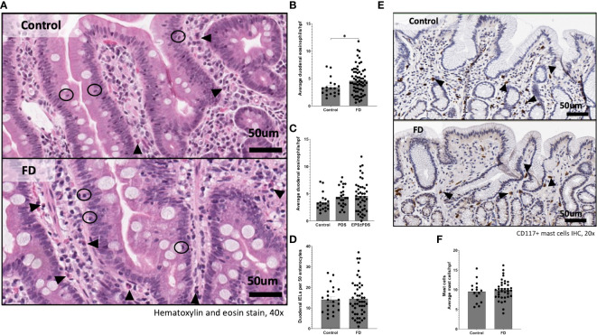
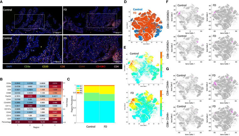
We identified duodenal T cell populations from 23 controls and 49 Rome III FD patients by flow cytometry using a surface marker antibody panel. We also analysed T cell populations in peripheral blood from 37 controls and 61 patients. Where available, we examined the number of duodenal eosinophils in patients and controls.
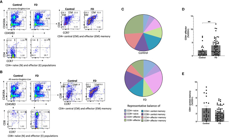
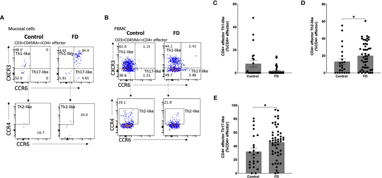
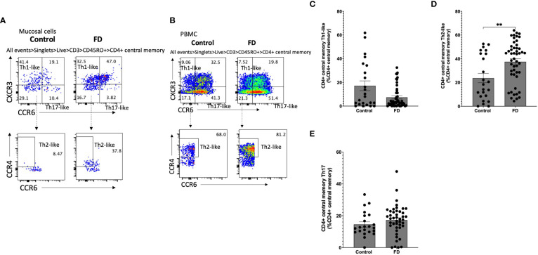
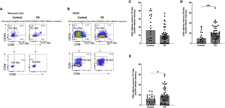
There was a shift in the duodenal T helper cell balance in FD patients compared to controls. For example, patients had increased duodenal mucosal Th2 populations in the effector (13.03 ± 16.11, 19.84 ± 15.51, =0.038), central memory (23.75 ± 18.97, 37.52 ± 17.51, =0.007) and effector memory (9.80±10.50 vs 20.53±14.15, =0.001) populations. Th17 populations were also increased in the effector (31.74±24.73 vs 45.57±23.75, =0.03) and effector memory (11.95±8.42 vs 18.44±15.63, =0.027) subsets. Peripheral T cell populations were unchanged between FD and control.
Our findings identify an association between lymphocyte populations and FD, specifically a Th2 and Th17 signature in the duodenal mucosa. The presence of effector and memory cells suggest that the microinflammation in FD is antigen driven.
功能性消化不良的特征是餐后不适或上腹痛的慢性症状,与明确的结构病理学无关。先前在患者中发现外周肠道归巢 T 细胞增加。迄今为止,尚不清楚这些 T 细胞是否具有抗原经验,或者是否与 FD 相关的特定表型。
本研究旨在描述 FD 患者血液和十二指肠黏膜中的 T 细胞群体,这些细胞可能与疾病发病机制有关。
我们通过流式细胞术使用表面标记抗体谱从 23 名对照和 49 名罗马 III FD 患者中鉴定十二指肠 T 细胞群体。我们还分析了 37 名对照和 61 名患者外周血中的 T 细胞群体。在有条件的情况下,我们检查了患者和对照者十二指肠嗜酸性粒细胞的数量。
与对照组相比,FD 患者十二指肠辅助性 T 细胞平衡发生了变化。例如,患者十二指肠黏膜 Th2 群体增加,效应细胞(13.03 ± 16.11,19.84 ± 15.51,=0.038)、中央记忆(23.75 ± 18.97,37.52 ± 17.51,=0.007)和效应记忆(9.80±10.50 vs 20.53±14.15,=0.001)。效应细胞(31.74±24.73 vs 45.57±23.75,=0.03)和效应记忆(11.95±8.42 vs 18.44±15.63,=0.027)亚群中 Th17 群体也增加。FD 和对照之间外周 T 细胞群体无变化。
我们的发现表明淋巴细胞群体与 FD 之间存在关联,特别是十二指肠黏膜中的 Th2 和 Th17 特征。效应和记忆细胞的存在表明 FD 中的微炎症是抗原驱动的。