Institute of Pathology, Philipps University of Marburg and University Hospital Giessen and Marburg GmbH, Marburg, Germany.
Department of Gynecology and Obstetrics, Breast Center Regio, Philipps University of Marburg and University Hospital Giessen and Marburg GmbH, Marburg, Germany.
PLoS One. 2023 Jan 24;18(1):e0280936. doi: 10.1371/journal.pone.0280936. eCollection 2023.
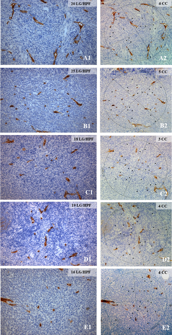
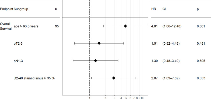
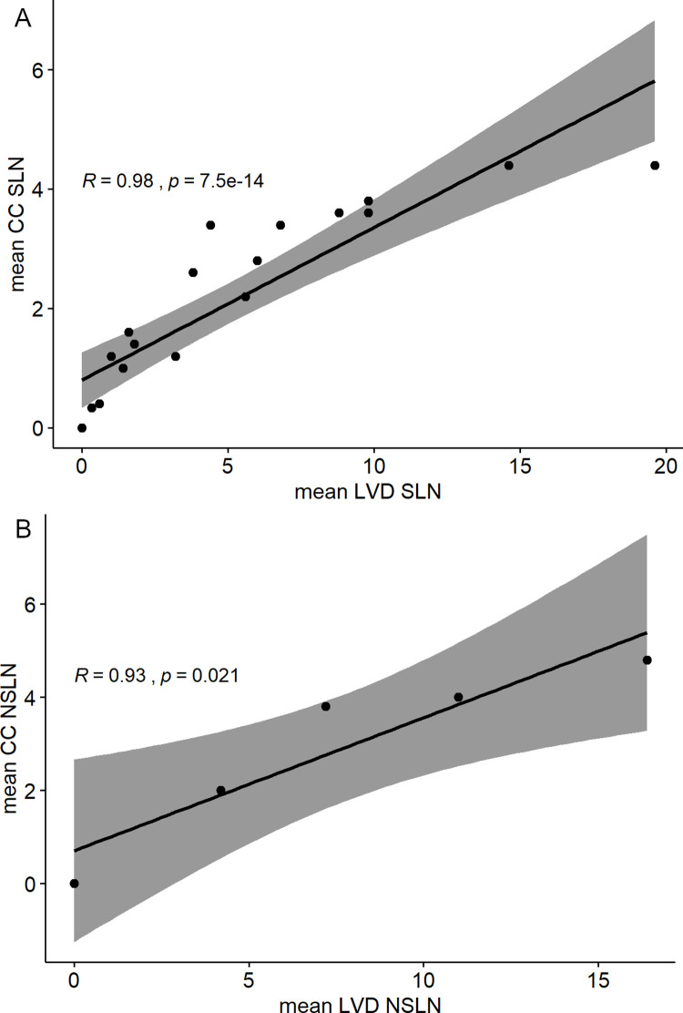
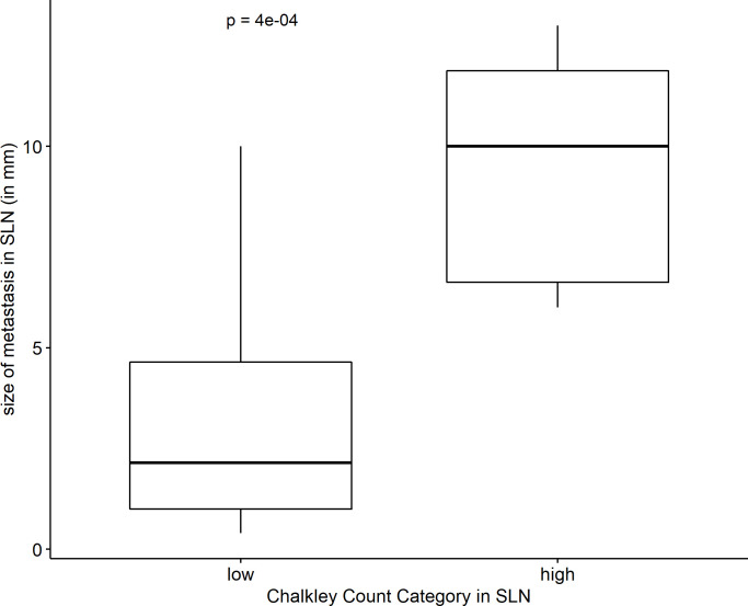
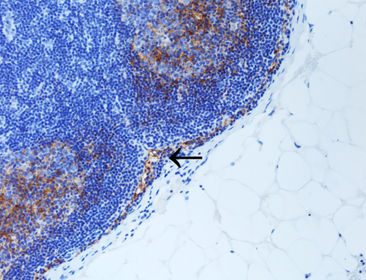
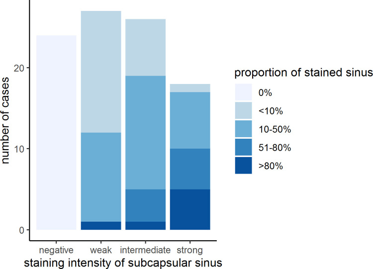
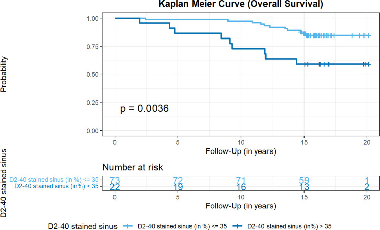
Several studies have demonstrated the de novo formation of lymphatic vessels or the reorganization of lymphatic sinus in tumor-draining lymph nodes, partly preceding the detection of lymphatic metastases. This "lymphovascular niche"is supposed to facilitate the survival of metastatic tumor cells. Few studies on nodal lymphangiogenesis in invasive breast cancer (BC) have been published, not considering tumor-free sentinel lymph nodes (SLN) and tumor types. Specimens of SLN and/ or non-SLN (NSLN) of 95 patients with BC were examined immunohistochemically for expression of the lymphatic endothelial marker D2-40 (podoplanin) on lymphatic vessels and the subcapsular sinus. The number of D2-40-positive lymph vessels in metastases was evaluated with two morphometric methods (Chalkley count and number per HPF). Data was explored with respect to TNM parameters, grading, tumor type, size of metastasis, lymph vessel number and hormone receptor/HER2 status with appropriate statistical tests. Lymphangiogenesis was detected exclusively in and around BC metastases with both methods for lymph vessel quantification being equivalent. Lymph vessel number correlated with the size of metastases, being significantly higher in larger metastases (p < 0.001). There was no significant statistical difference with respect to tumor types. Intranodal lymphangiogenesis could not be verified by D2-40 staining in any of the tumor-free lymph nodes examined. However, D2-40 was frequently detected in sinus endothelial/virgultar cells of the subcapsular sinus, partly with strong uniform positivity. Staining intensity and stained proportion of the subcapsular sinus were markedly heterogeneous, significantly correlating with each other both in SLN and NSLN (p < 0.001). A higher proportion of D2-40 stained subcapsular sinus in SLN was significantly associated with worse overall survival (p = 0.0036) and an independent prognostic parameter in multivariate analysis (p = 0.033, HR 2.87). Further studies are necessary to elucidate the biological and clinical significance of the observed immunophenotypic variations of nodal sinus endothelium.
已有多项研究证实,在肿瘤引流淋巴结中,新生淋巴管的形成或淋巴管窦的再组织化部分先于淋巴转移的检测。这个“淋巴血管龛”被认为有助于转移性肿瘤细胞的存活。关于浸润性乳腺癌(BC)中淋巴结淋巴管生成的研究较少,且这些研究没有考虑到无肿瘤的前哨淋巴结(SLN)和肿瘤类型。对 95 例 BC 患者的 SLN 和/或非 SLN(NSLN)标本进行免疫组织化学检查,检测淋巴管内皮标志物 D2-40(足细胞蛋白)的表达和被膜下窦。采用两种形态计量学方法(查尔克利计数和每高倍镜视野数)评估转移灶中 D2-40 阳性淋巴管的数量。通过适当的统计检验,根据 TNM 参数、分级、肿瘤类型、转移灶大小、淋巴管数量和激素受体/HER2 状态对数据进行了探讨。淋巴生成仅在 BC 转移灶内及其周围被检测到,两种淋巴管定量方法的结果相当。淋巴管数量与转移灶大小相关,在较大的转移灶中显著更高(p<0.001)。肿瘤类型之间无显著统计学差异。在任何检查的无肿瘤淋巴结中,都不能通过 D2-40 染色来验证淋巴结内的淋巴管生成。然而,D2-40 经常在被膜下窦的窦内皮/间充质细胞中被检测到,部分呈强均匀阳性。被膜下窦的染色强度和染色比例明显不均匀,在 SLN 和 NSLN 中彼此显著相关(p<0.001)。SLN 中 D2-40 染色的被膜下窦比例较高与总生存时间更差显著相关(p=0.0036),并且在多变量分析中是独立的预后参数(p=0.033,HR 2.87)。需要进一步的研究来阐明观察到的淋巴结窦内皮免疫表型变化的生物学和临床意义。