Center for Neurobiology and Vaccine Development, Ophthalmology Research, Department of Surgery, Cedars-Sinai Medical Center, Los Angeles, CA, USA.
Applied Genomics, Computation, and Translational Core, Cedars-Sinai Medical Center, Los Angeles, CA, USA.
Sci Adv. 2023 Jan 25;9(4):eadf4904. doi: 10.1126/sciadv.adf4904.
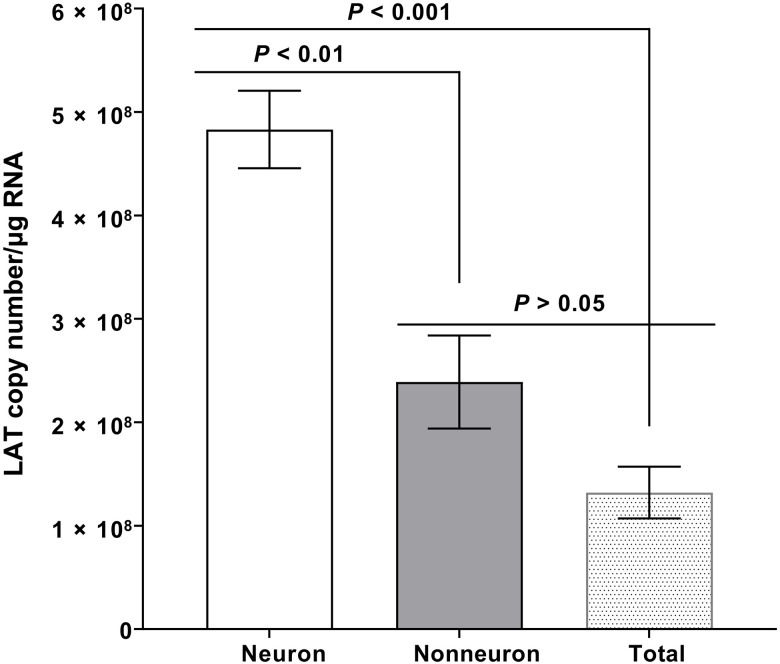
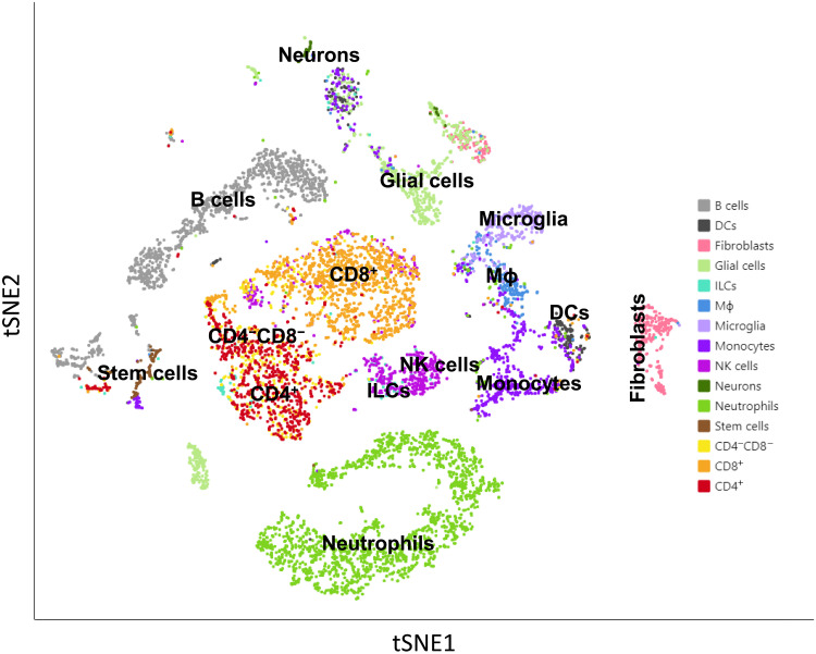
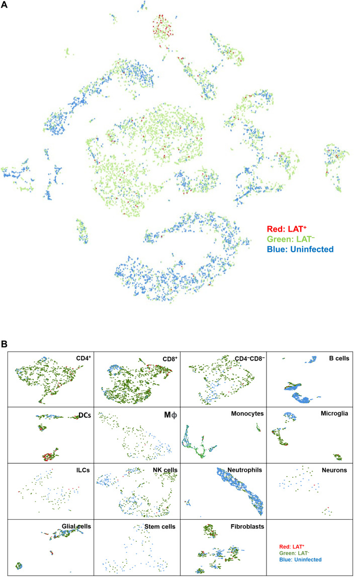
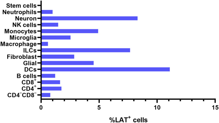
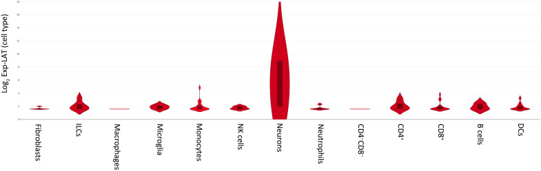
A hallmark of herpes simplex virus (HSV) infection is the establishment of latent virus in peripheral sensory ganglia of the latently infected host. We and others originally reported that the latency-associated transcript (LAT) is the only abundantly expressed viral gene in neurons within trigeminal ganglia (TG) of a latently infected host. Here, we investigated the possible contribution of various cells [i.e., B cells, dendritic cells (DCs), fibroblasts, glial cells, innate lymphoid cells (ILCs), macrophages, microglia, monocytes, natural killer cells, neurons, neutrophils, and T cells] isolated from TG of latently infected mice. Our results demonstrated that all of these cell types contain LAT, with DCs, neurons, and ILCs having the most LAT cells. These results suggest that HSV-1 can establish a quiescent/latent infection in a subset of nonneuronal cells, which enhances the chances that the virus will survive in its host.
单纯疱疹病毒 (HSV) 感染的一个标志是潜伏病毒在潜伏感染宿主周围感觉神经节中的建立。我们和其他人最初报道潜伏相关转录物 (LAT) 是潜伏感染宿主三叉神经节 (TG) 内神经元中唯一大量表达的病毒基因。在这里,我们研究了从潜伏感染小鼠 TG 中分离的各种细胞[即 B 细胞、树突状细胞 (DCs)、成纤维细胞、神经胶质细胞、固有淋巴细胞 (ILCs)、巨噬细胞、小胶质细胞、单核细胞、自然杀伤细胞、神经元、嗜中性粒细胞和 T 细胞]可能做出的贡献。我们的结果表明,所有这些细胞类型都含有 LAT,其中 DCs、神经元和 ILCs 含有最多的 LAT 细胞。这些结果表明,HSV-1 可以在一组非神经元细胞中建立静止/潜伏感染,这增加了病毒在宿主体内存活的机会。