• 文献检索
  • 文档翻译
  • 深度研究
  • 学术资讯
  • Suppr Zotero 插件Zotero 插件
  • 邀请有礼
  • 套餐&价格
  • 历史记录
应用&插件
Suppr Zotero 插件Zotero 插件浏览器插件Mac 客户端Windows 客户端微信小程序
定价
高级版会员购买积分包购买API积分包
服务
文献检索文档翻译深度研究API 文档MCP 服务
关于我们
关于 Suppr公司介绍联系我们用户协议隐私条款
关注我们

Suppr 超能文献

核心技术专利:CN118964589B侵权必究
粤ICP备2023148730 号-1Suppr @ 2026

文献检索

告别复杂PubMed语法,用中文像聊天一样搜索,搜遍4000万医学文献。AI智能推荐,让科研检索更轻松。

立即免费搜索

文件翻译

保留排版,准确专业,支持PDF/Word/PPT等文件格式,支持 12+语言互译。

免费翻译文档

深度研究

AI帮你快速写综述,25分钟生成高质量综述,智能提取关键信息,辅助科研写作。

立即免费体验

原代细胞二维培养中的骨形成

Bone Formation in 2D Culture of Primary Cells.

作者信息

Mertz Edward L, Makareeva Elena, Mirigian Lynn S, Leikin Sergey

机构信息

Eunice Kennedy Shriver National Institute of Health and Human Development National Institutes of Health Bethesda MD USA.

出版信息

JBMR Plus. 2022 Dec 13;7(1):e10701. doi: 10.1002/jbm4.10701. eCollection 2023 Jan.

DOI:10.1002/jbm4.10701
PMID:36699640
原文链接:https://pmc.ncbi.nlm.nih.gov/articles/PMC9850442/
Abstract

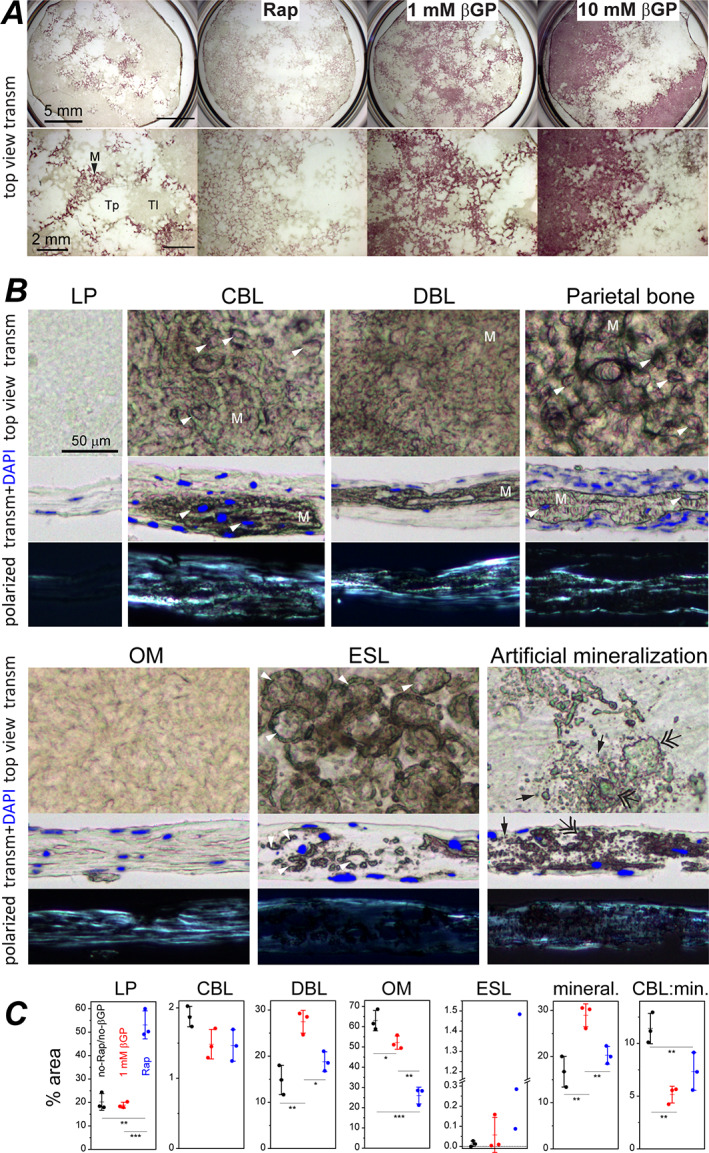
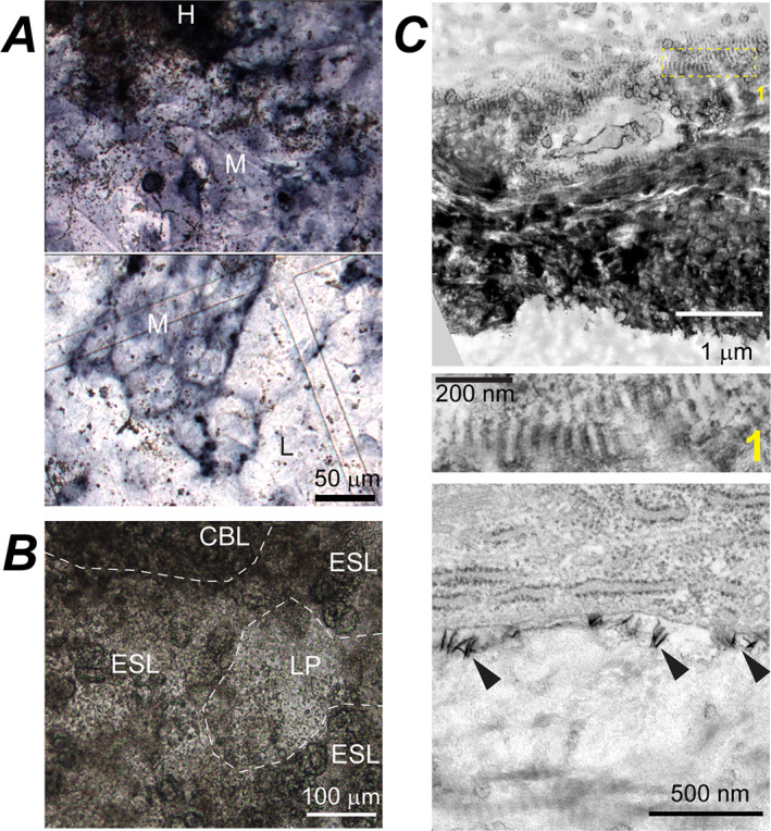
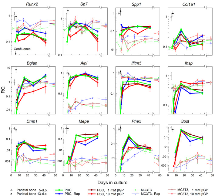
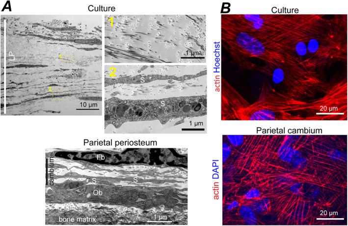
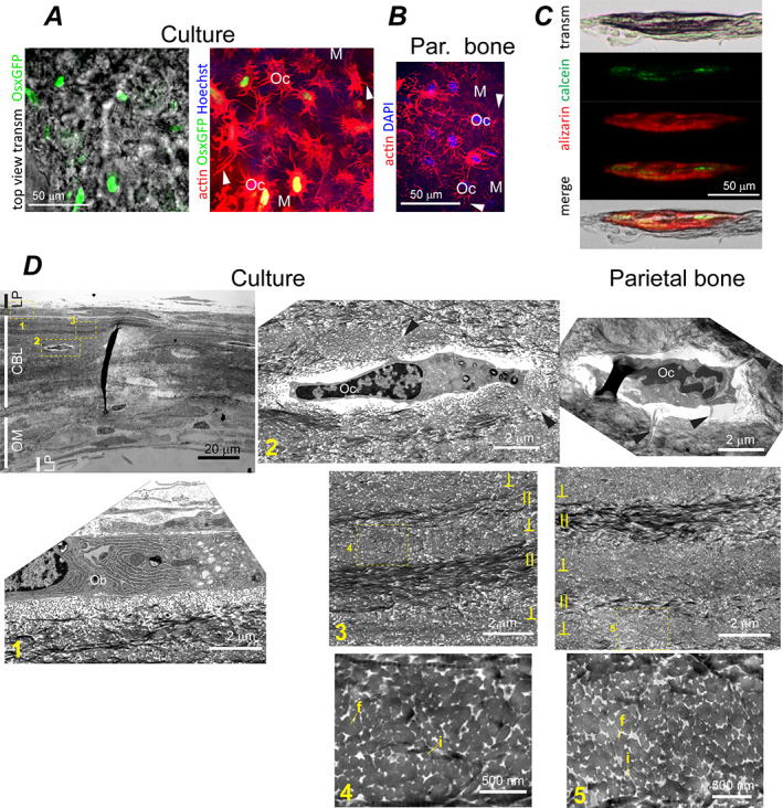
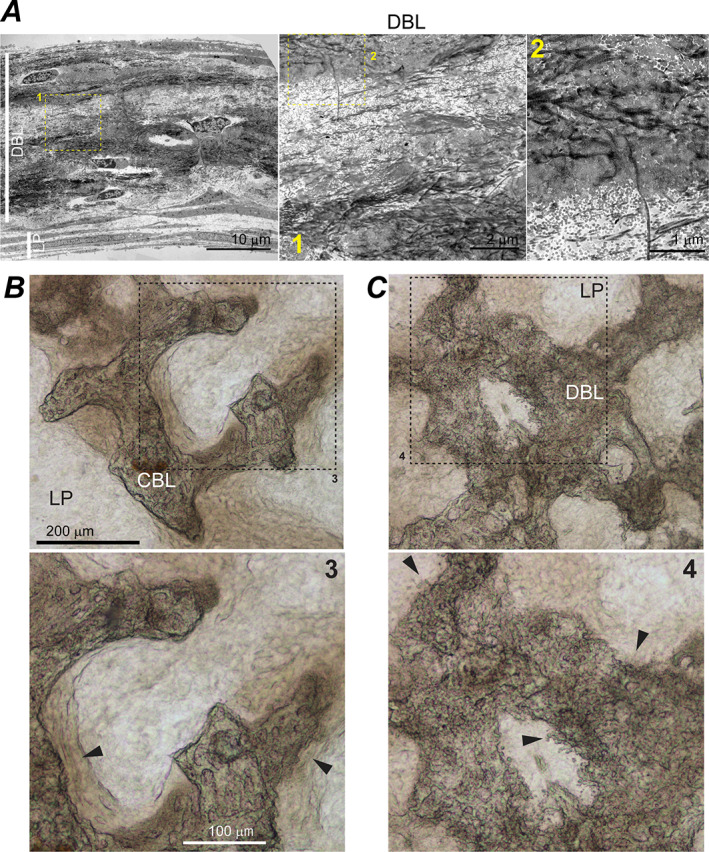
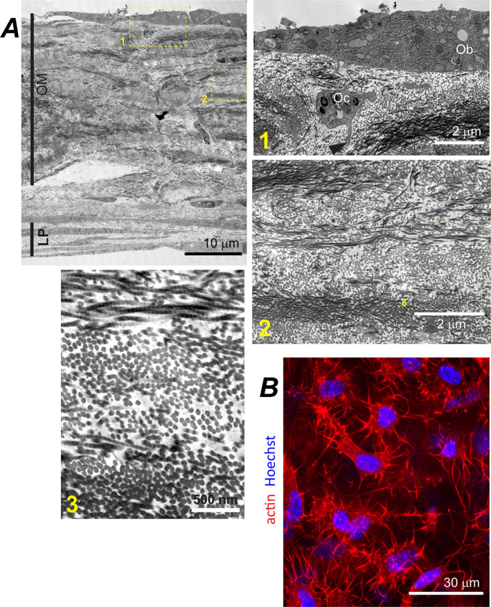
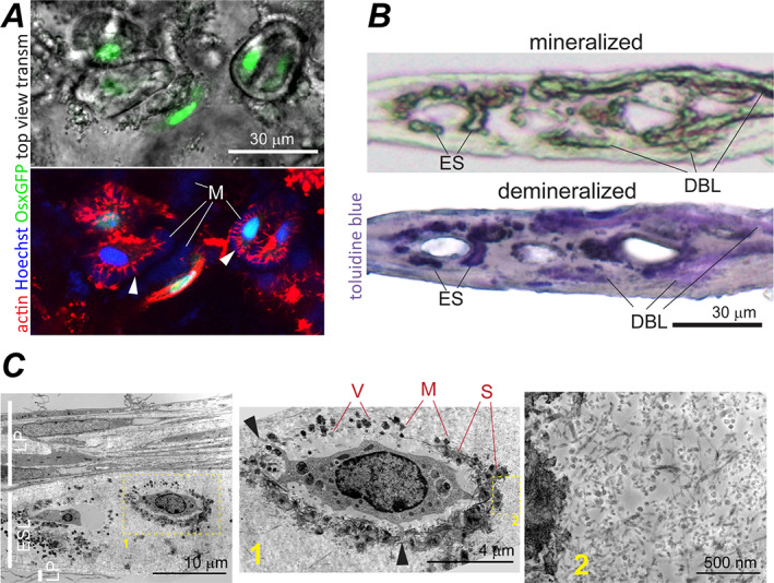
Relevance of mineralized nodules in two-dimensional (2D) osteoblast/osteocyte cultures to bone biology, pathology, and engineering is a decades old question, but a comprehensive answer appears to be still wanting. Bone-like cells, extracellular matrix (ECM), and mineral were all reported but so were non-bone-like ones. Many studies described seemingly bone-like cell-ECM structures based on similarity to few select bone features in vivo, yet no studies examined multiple bone features simultaneously and none systematically studied all types of structures coexisting in the same culture. Here, we report such comprehensive analysis of 2D cultures based on light and electron microscopies, Raman microspectroscopy, gene expression, and in situ messenger RNA (mRNA) hybridization. We demonstrate that 2D cultures of primary cells from mouse calvaria do form bona fide bone. Cells, ECM, and mineral within it exhibit morphology, structure, ultrastructure, composition, spatial-temporal gene expression pattern, and growth consistent with intramembranous ossification. However, this bone is just one of at least five different types of cell-ECM structures coexisting in the same 2D culture, which vary widely in their resemblance to bone and ability to mineralize. We show that the other two mineralizing structures may represent abnormal (disrupted) bone and cartilage-like structure with chondrocyte-to-osteoblast transdifferentiation. The two nonmineralizing cell-ECM structures may mimic periosteal cambium and pathological, nonmineralizing osteoid. Importantly, the most commonly used culture conditions (10mM β-glycerophosphate) induce artificial mineralization of all cell-ECM structures, which then become barely distinguishable. We therefore discuss conditions and approaches promoting formation of bona fide bone and simple means for distinguishing it from the other cell-ECM structures. Our findings may improve osteoblast differentiation and function analyses based on 2D cultures and extend applications of these cultures to general bone biology and tissue engineering research. Published 2022. This article is a U.S. Government work and is in the public domain in the USA. published by Wiley Periodicals LLC on behalf of American Society for Bone and Mineral Research.

摘要

二维(2D)成骨细胞/骨细胞培养中矿化结节与骨生物学、病理学和工程学的相关性是一个存在了数十年的问题,但似乎仍缺乏全面的答案。已报道了类骨细胞、细胞外基质(ECM)和矿物质,但也有非类骨细胞的报道。许多研究基于与体内少数选定骨特征的相似性描述了看似类骨的细胞-ECM结构,但没有研究同时检查多个骨特征,也没有系统地研究同一培养物中共存的所有类型的结构。在这里,我们报告基于光学和电子显微镜、拉曼显微光谱、基因表达和原位信使核糖核酸(mRNA)杂交对2D培养物进行的这种全面分析。我们证明,来自小鼠颅骨的原代细胞的2D培养确实形成了真正的骨。其中的细胞、ECM和矿物质表现出与膜内成骨一致的形态、结构、超微结构、组成、时空基因表达模式和生长。然而,这种骨只是同一2D培养物中共存的至少五种不同类型的细胞-ECM结构之一,它们在与骨的相似性和矿化能力方面差异很大。我们表明,其他两种矿化结构可能代表具有软骨细胞向成骨细胞转分化的异常(破坏的)骨和软骨样结构。两种非矿化细胞-ECM结构可能模拟骨膜形成层和病理性、非矿化类骨质。重要的是,最常用的培养条件(10mMβ-甘油磷酸)会诱导所有细胞-ECM结构的人工矿化,然后它们变得几乎无法区分。因此,我们讨论了促进真正骨形成的条件和方法,以及将其与其他细胞-ECM结构区分开来的简单方法。我们的发现可能会改善基于2D培养的成骨细胞分化和功能分析,并将这些培养物的应用扩展到一般骨生物学和组织工程研究。发表于2022年。本文是美国政府的作品,在美国属于公共领域。由Wiley Periodicals LLC代表美国骨与矿物质研究学会发表。

https://cdn.ncbi.nlm.nih.gov/pmc/blobs/36ec/9850442/cd8f2482a752/JBM4-7-e10701-g004.jpg
https://cdn.ncbi.nlm.nih.gov/pmc/blobs/36ec/9850442/7984d85f4aeb/JBM4-7-e10701-g010.jpg
https://cdn.ncbi.nlm.nih.gov/pmc/blobs/36ec/9850442/789d0123eaf5/JBM4-7-e10701-g008.jpg
https://cdn.ncbi.nlm.nih.gov/pmc/blobs/36ec/9850442/bd6c7303bc54/JBM4-7-e10701-g009.jpg
https://cdn.ncbi.nlm.nih.gov/pmc/blobs/36ec/9850442/5a8b8508dc49/JBM4-7-e10701-g003.jpg
https://cdn.ncbi.nlm.nih.gov/pmc/blobs/36ec/9850442/6fc5b84b1ba7/JBM4-7-e10701-g011.jpg
https://cdn.ncbi.nlm.nih.gov/pmc/blobs/36ec/9850442/68a422017f12/JBM4-7-e10701-g007.jpg
https://cdn.ncbi.nlm.nih.gov/pmc/blobs/36ec/9850442/5fc3caa93395/JBM4-7-e10701-g006.jpg
https://cdn.ncbi.nlm.nih.gov/pmc/blobs/36ec/9850442/b142965b7466/JBM4-7-e10701-g001.jpg
https://cdn.ncbi.nlm.nih.gov/pmc/blobs/36ec/9850442/5b6af639739e/JBM4-7-e10701-g002.jpg
https://cdn.ncbi.nlm.nih.gov/pmc/blobs/36ec/9850442/cd8f2482a752/JBM4-7-e10701-g004.jpg

相似文献

1
Bone Formation in 2D Culture of Primary Cells.原代细胞二维培养中的骨形成
JBMR Plus. 2022 Dec 13;7(1):e10701. doi: 10.1002/jbm4.10701. eCollection 2023 Jan.
2
Type VI Collagen Regulates Endochondral Ossification in the Temporomandibular Joint.VI型胶原蛋白调节颞下颌关节的软骨内成骨。
JBMR Plus. 2022 Mar 10;6(5):e10617. doi: 10.1002/jbm4.10617. eCollection 2022 May.
3
Extracellular matrix mineralization in murine MC3T3-E1 osteoblast cultures: an ultrastructural, compositional and comparative analysis with mouse bone.小鼠MC3T3-E1成骨细胞培养中的细胞外基质矿化:与小鼠骨骼的超微结构、成分及比较分析
Bone. 2015 Feb;71:244-56. doi: 10.1016/j.bone.2014.11.003. Epub 2014 Nov 13.
4
Osteocyte-Like Cells Differentiated From Primary Osteoblasts in an Artificial Human Bone Tissue Model.在人工构建的人体骨组织模型中,由原代成骨细胞分化而来的类骨细胞。
JBMR Plus. 2023 Jun 28;7(9):e10792. doi: 10.1002/jbm4.10792. eCollection 2023 Sep.
5
In situ examination of osteoblast biomineralization on sulfonated polystyrene-modified substrates using Fourier transform infrared microspectroscopy.使用傅里叶变换红外显微光谱法对磺化聚苯乙烯改性基质上的成骨细胞生物矿化进行原位检测。
Biointerphases. 2017 Jul 10;12(3):031001. doi: 10.1116/1.4992137.
6
Time lapse imaging techniques for comparison of mineralization dynamics in primary murine osteoblasts and the late osteoblast/early osteocyte-like cell line MLO-A5.用于比较原代小鼠成骨细胞和晚期成骨细胞/早期骨细胞样细胞系MLO-A5矿化动力学的延时成像技术。
Cells Tissues Organs. 2009;189(1-4):6-11. doi: 10.1159/000151745. Epub 2008 Aug 27.
7
Expression of extracellular matrix proteins in human periodontal ligament cells during mineralization in vitro.体外矿化过程中人类牙周膜细胞外基质蛋白的表达
J Periodontol. 1997 Apr;68(4):320-7. doi: 10.1902/jop.1997.68.4.320.
8
Extracellular matrix mineralization promotes E11/gp38 glycoprotein expression and drives osteocytic differentiation.细胞外基质矿化促进 E11/gp38 糖蛋白表达并驱动破骨细胞分化。
PLoS One. 2012;7(5):e36786. doi: 10.1371/journal.pone.0036786. Epub 2012 May 7.
9
A Novel Osteogenic Cell Line That Differentiates Into GFP-Tagged Osteocytes and Forms Mineral With a Bone-Like Lacunocanalicular Structure.一种新型成骨细胞系,可分化为 GFP 标记的成骨细胞,并形成具有类似骨陷窝和小管结构的矿化物质。
J Bone Miner Res. 2019 Jun;34(6):979-995. doi: 10.1002/jbmr.3720. Epub 2019 Jun 7.
10
Solid-State Nuclear Magnetic Resonance (SSNMR) Characterization of Osteoblasts From Mesenchymal Stromal Cell Differentiation to Osteoblast Mineralization.从间充质基质细胞分化为成骨细胞并矿化过程中,成骨细胞的固态核磁共振(SSNMR)表征
JBMR Plus. 2022 Sep 12;6(10):e10662. doi: 10.1002/jbm4.10662. eCollection 2022 Oct.

引用本文的文献

1
Continuous pore size gradient enhances zonal-specific differentiation of stem cells in an osteochondral scaffold.连续孔径梯度增强了骨软骨支架中干细胞的区域特异性分化。
RSC Adv. 2025 Aug 11;15(35):28452-28463. doi: 10.1039/d5ra00540j.
2
Modeling methods of different tumor organoids and their application in tumor drug resistance research.不同肿瘤类器官的建模方法及其在肿瘤耐药性研究中的应用。
Cancer Drug Resist. 2025 Jul 1;8:32. doi: 10.20517/cdr.2025.34. eCollection 2025.
3
Long-Term Culture of Cellular Spheroids in Novel Hydrogel Constructs for ECM Characterization in Bone Models.

本文引用的文献

1
Noncanonical ER-Golgi trafficking and autophagy of endogenous procollagen in osteoblasts.成骨细胞中内源性原胶原的非典型内质网-高尔基体运输和自噬。
Cell Mol Life Sci. 2021 Dec;78(24):8283-8300. doi: 10.1007/s00018-021-04017-z. Epub 2021 Nov 15.
2
Gli1 progenitors mediate bone anabolic function of teriparatide via Hh and Igf signaling.Gli1 祖细胞通过 HH 和 IGF 信号介导特立帕肽的骨合成作用。
Cell Rep. 2021 Aug 17;36(7):109542. doi: 10.1016/j.celrep.2021.109542.
3
Evaluation of MC3T3-E1 Cell Osteogenesis in Different Cell Culture Media.
用于骨模型中细胞外基质表征的新型水凝胶构建体中细胞球体的长期培养
Materials (Basel). 2025 Jul 28;18(15):3538. doi: 10.3390/ma18153538.
4
Osteoclast-derived coupling factors: origins and state-of-play Louis V Avioli lecture, ASBMR 2023.破骨细胞衍生的偶联因子:起源和现状 路易·V·阿维奥利讲座,ASBMR 2023 年。
J Bone Miner Res. 2024 Sep 26;39(10):1377-1385. doi: 10.1093/jbmr/zjae110.
5
RNA-based bone histomorphometry: method and its application to explaining postpubertal bone gain in a G610C mouse model of osteogenesis imperfecta.基于 RNA 的骨组织形态计量学:方法及其在解释成骨不全症 G610C 小鼠模型中青春期后骨量增加的应用。
J Bone Miner Res. 2024 Mar 22;39(2):177-189. doi: 10.1093/jbmr/zjad004.
不同细胞培养液中 MC3T3-E1 细胞成骨的评价。
Int J Mol Sci. 2021 Jul 20;22(14):7752. doi: 10.3390/ijms22147752.
4
Mechanisms of procollagen and HSP47 sorting during ER-to-Golgi trafficking.胶原前体和 HSP47 在 ER 到高尔基体运输过程中的分拣机制。
Matrix Biol. 2020 Nov;93:79-94. doi: 10.1016/j.matbio.2020.06.002. Epub 2020 Jun 17.
5
Alkaline phosphatase: Structure, expression and its function in bone mineralization.碱性磷酸酶:结构、表达及其在骨矿化中的作用
Gene. 2020 Sep 5;754:144855. doi: 10.1016/j.gene.2020.144855. Epub 2020 Jun 6.
6
Models of Bone Remodelling and Associated Disorders.骨重塑及相关疾病模型
Front Bioeng Biotechnol. 2018 Oct 11;6:134. doi: 10.3389/fbioe.2018.00134. eCollection 2018.
7
Noncanonical autophagy at ER exit sites regulates procollagen turnover.内质网出口位点的非典型自噬调节原胶原的周转。
Proc Natl Acad Sci U S A. 2018 Oct 23;115(43):E10099-E10108. doi: 10.1073/pnas.1814552115. Epub 2018 Oct 4.
8
Bone remodelling in vitro: Where are we headed?: -A review on the current understanding of physiological bone remodelling and inflammation and the strategies for testing biomaterials in vitro.体外骨重建:我们的方向在哪里?——对生理骨重建和炎症的现有理解的综述,以及体外测试生物材料的策略。
Bone. 2018 May;110:38-46. doi: 10.1016/j.bone.2018.01.015. Epub 2018 Feb 3.
9
Bone Cell Models: Impact of Fluid Shear Stress on Bone Formation.骨细胞模型:流体剪切应力对骨形成的影响。
Front Bioeng Biotechnol. 2016 Nov 15;4:87. doi: 10.3389/fbioe.2016.00087. eCollection 2016.
10
Supramolecular Organization of Collagen Fibrils in Healthy and Osteoarthritic Human Knee and Hip Joint Cartilage.健康和骨关节炎患者膝关节及髋关节软骨中胶原纤维的超分子结构
PLoS One. 2016 Oct 25;11(10):e0163552. doi: 10.1371/journal.pone.0163552. eCollection 2016.