Li Li, Nguyen Binh, Mullapudi Vishruth, Saelices Lorena, Joachimiak Lukasz A
Center for Alzheimer's and Neurodegenerative Diseases, Peter O'Donnell Jr. Brain Institute, University of Texas Southwestern Medical Center, Dallas, Texas 75390.
Department of Biophysics, University of Texas Southwestern Medical Center, Dallas, Texas 75390.
bioRxiv. 2023 Jan 10:2023.01.10.523459. doi: 10.1101/2023.01.10.523459.
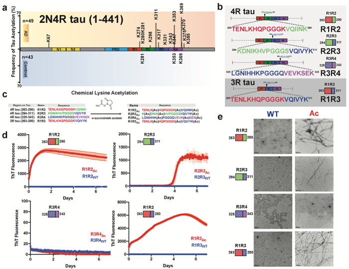
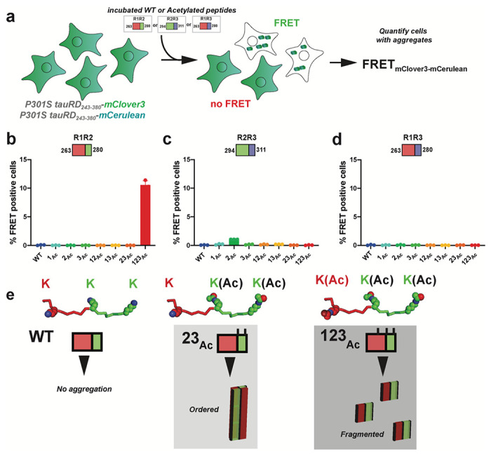
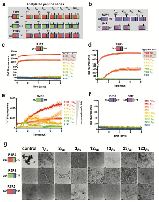
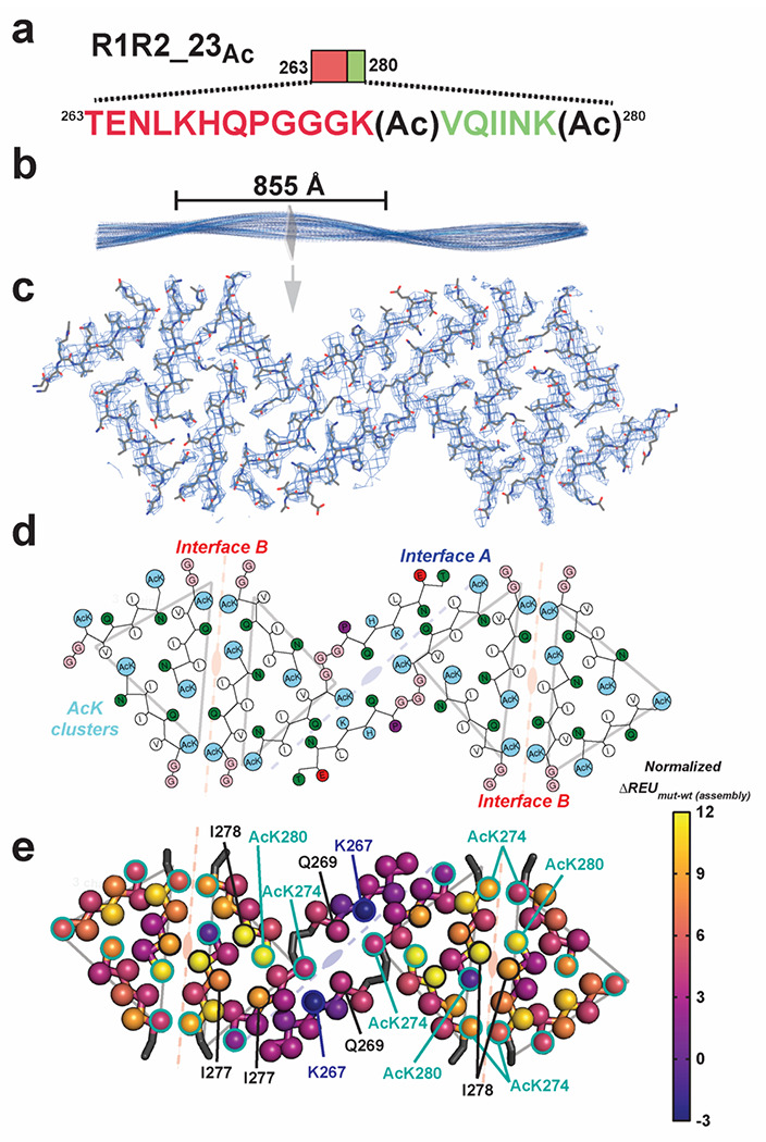
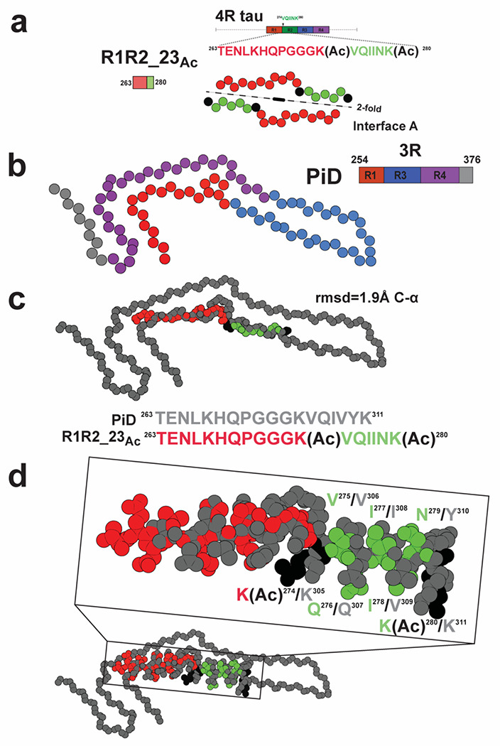
Assembly of the microtubule-associated protein into tauopathy fibril conformations dictates the pathology of a diversity of diseases. Recent cryogenic Electron Microscopy (cryo-EM) structures have uncovered distinct fibril conformations in different tauopathies but it remains unknown how these structures fold from a single protein sequence. It has been proposed that post-translational modifications may drive tau assembly but no direct mechanism for how modifications drive assembly has emerged. Leveraging established aggregation-regulating tau fragments that are normally inert, we tested the effect of chemical modification of lysines with acetyl groups on tau fragment conversion into amyloid aggregates. We identify specific patterns of acetylation that flank amyloidogenic motifs on the tau fragments that drive rapid fibril assembly. To understand how this pattern of acetylation may drive assembly, we determined a 3.9 Å cryo-EM structure of an amyloid fibril assembled from an acetylated tau fragment. The structure uncovers how lysine acetylation patterns mediate gain-of-function interactions to promote amyloid assembly. Comparison of the structure to an ex vivo tau fibril conformation from Pick's Disease reveals regions of high structural similarity. Finally, we show that our lysine- acetylated sequences exhibit fibril assembly activity in cell-based tau aggregation assays. Our data uncover the dual role of lysine residues in limiting aggregation while their acetylation leads to stabilizing pro-aggregation interactions. Design of tau sequence with specific acetylation patterns may lead to controllable tau aggregation to direct folding of tau into distinct folds.
微管相关蛋白组装成tau蛋白病原纤维构象决定了多种疾病的病理。最近的低温电子显微镜(cryo-EM)结构揭示了不同tau蛋白病中独特的原纤维构象,但这些结构如何从单一蛋白质序列折叠而成仍不清楚。有人提出翻译后修饰可能驱动tau蛋白组装,但尚未出现修饰如何驱动组装的直接机制。利用已建立的通常无活性的聚集调节tau片段,我们测试了用乙酰基化学修饰赖氨酸对tau片段转化为淀粉样聚集体的影响。我们确定了tau片段上淀粉样生成基序两侧的特定乙酰化模式,这些模式驱动快速的原纤维组装。为了理解这种乙酰化模式如何驱动组装,我们确定了由乙酰化tau片段组装而成的淀粉样原纤维的3.9埃低温电子显微镜结构。该结构揭示了赖氨酸乙酰化模式如何介导功能获得性相互作用以促进淀粉样组装。将该结构与来自匹克病的体外tau原纤维构象进行比较,发现了高度结构相似的区域。最后,我们表明我们的赖氨酸乙酰化序列在基于细胞的tau聚集测定中表现出原纤维组装活性。我们的数据揭示了赖氨酸残基在限制聚集方面的双重作用,而它们的乙酰化导致稳定促聚集相互作用。设计具有特定乙酰化模式的tau序列可能导致可控的tau聚集,以指导tau折叠成不同的构象。