Shrestha Rabina, McCann Tess, Saravanan Harini, Lieberth Jaret, Koirala Prashanna, Bloomekatz Joshua
Department of Biology, University of Mississippi, University, MS 38677.
bioRxiv. 2023 Jan 3:2023.01.03.522612. doi: 10.1101/2023.01.03.522612.
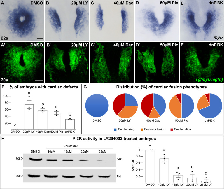
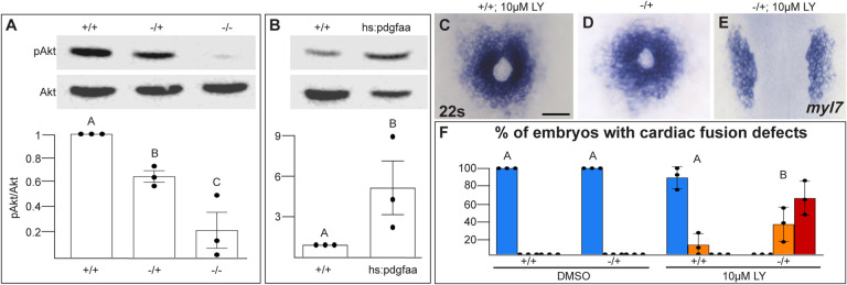
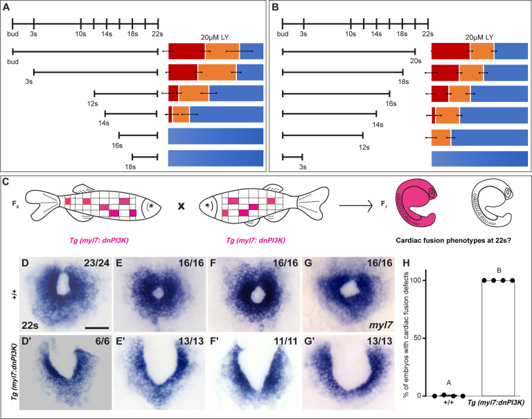
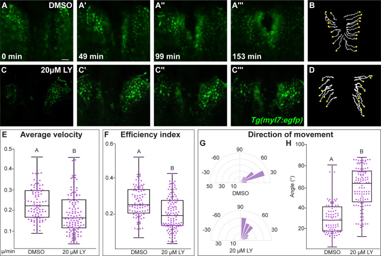
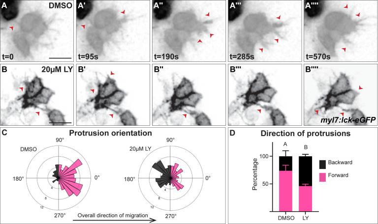
Coordinated cell movement is a fundamental process in organ formation. During heart development, bilateral myocardial precursors collectively move towards the midline (cardiac fusion) to form the primitive heart tube. Along with extrinsic influences such as the adjacent anterior endoderm which are known to be required for cardiac fusion, we previously showed that the platelet-derived growth factor receptor alpha (Pdgfra) is also required. However, an intrinsic mechanism that regulates myocardial movement remains to be elucidated. Here, we uncover an essential intrinsic role in the myocardium for the phosphoinositide 3-kinase (PI3K) intracellular signaling pathway in directing myocardial movement towards the midline. imaging reveals that in PI3K-inhibited zebrafish embryos myocardial movements are misdirected and slower, while midline-oriented dynamic myocardial membrane protrusions become unpolarized. Moreover, PI3K activity is dependent on and genetically interacts with Pdgfra to regulate myocardial movement. Together our findings reveal an intrinsic myocardial steering mechanism that responds to extrinsic cues during the initiation of cardiac development.
协调的细胞运动是器官形成过程中的一个基本过程。在心脏发育过程中,双侧心肌前体细胞共同向中线移动(心脏融合)以形成原始心管。除了已知对心脏融合必需的外在影响因素,如相邻的前内胚层外,我们之前还表明血小板衍生生长因子受体α(Pdgfra)也是必需的。然而,调节心肌运动的内在机制仍有待阐明。在这里,我们发现磷酸肌醇3激酶(PI3K)细胞内信号通路在指导心肌向中线移动方面在心肌中起着至关重要的内在作用。成像显示,在PI3K抑制的斑马鱼胚胎中,心肌运动方向错误且速度较慢,而朝向中线的动态心肌膜突起变得无极化。此外,PI3K活性依赖于Pdgfra并与其发生基因相互作用以调节心肌运动。我们的研究结果共同揭示了一种内在的心肌引导机制,该机制在心脏发育起始过程中对外在信号作出反应。