Schweer David, Anand Namrata, Anderson Abigail, McCorkle J Robert, Neupane Khaga, Nail Alexandra N, Harvey Brock, Hill Kristen S, Ueland Frederick, Richards Christopher, Kolesar Jill
Division of Gynecologic Oncology, Department of Obstetrics and Gynecology, College of Medicine, University of Kentucky, Lexington, KY, United States.
Markey Cancer Center, University of Kentucky, Lexington, KY, United States.
Front Oncol. 2023 Jan 11;12:1042730. doi: 10.3389/fonc.2022.1042730. eCollection 2022.
Ovarian cancer is a deadly female malignancy with a high rate of recurrent and chemotherapy-resistant disease. Tumor-associated macrophages (TAMs) are a significant component of the tumor microenvironment and include high levels of M2-protumor macrophages that promote chemoresistance and metastatic spread. M2 macrophages can be converted to M1 anti-tumor macrophages, representing a novel therapeutic approach. Vesicles engineered from M1 macrophages (MEVs) are a novel method for converting M2 macrophages to M1 phenotype-like macrophages.
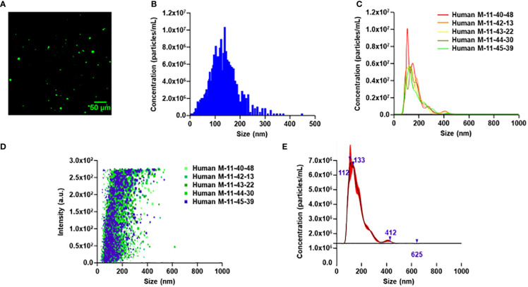
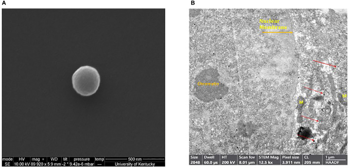
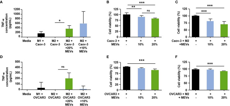
Macrophages were isolated and cultured from human peripheral blood mononuclear cells. Macrophages were stimulated to M1 or M2 phenotypes utilizing LPS/IFN-γ and IL-4/IL-13, respectively. M1 MEVs were generated with nitrogen cavitation and ultracentrifugation. Co-culture of ovarian cancer cells with macrophages and M1 MEVs was followed by cytokine, PCR, and cell viability analysis. Murine macrophage cell line, RAW264.7 cells were cultured and used to generate M1 MEVs for use in ovarian cancer xenograft models.
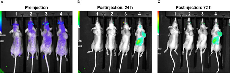
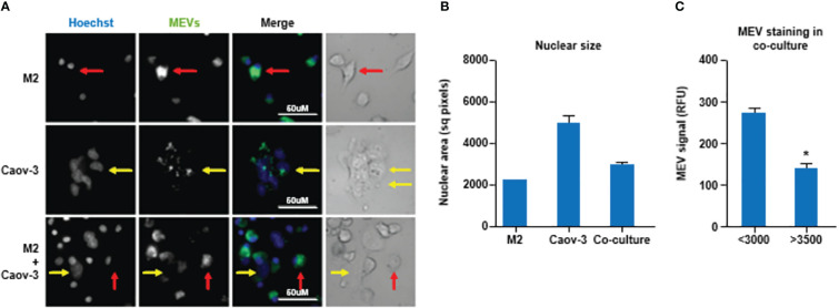
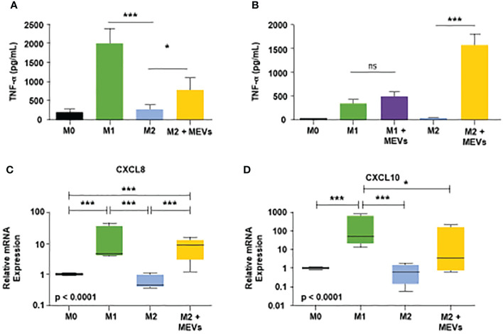
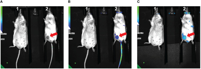
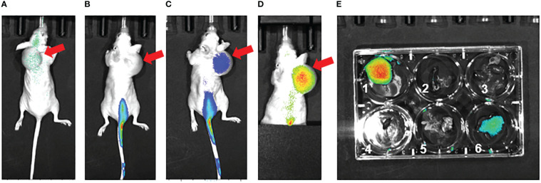
M1 MEVs can effectively convert M2 macrophages to an M1-like state both in isolation and when co-cultured with ovarian cancer cells , resulting in a reduced ovarian cancer cell viability. Additionally, RAW264.7 M1 MEVs can localize to ovarian cancer tumor xenografts in mice.
Human M1 MEVs can repolarize M2 macrophages to a M1 state and have anti-cancer activity against ovarian cancer cell lines. RAW264.7 M1 MEVs localize to tumor xenografts murine models.
卵巢癌是一种致命的女性恶性肿瘤,复发率和化疗耐药率很高。肿瘤相关巨噬细胞(TAM)是肿瘤微环境的重要组成部分,其中高水平的M2促肿瘤巨噬细胞会促进化疗耐药和转移扩散。M2巨噬细胞可转化为M1抗肿瘤巨噬细胞,这代表了一种新的治疗方法。由M1巨噬细胞工程化改造的囊泡(MEV)是将M2巨噬细胞转化为M1表型样巨噬细胞的新方法。
从人外周血单核细胞中分离并培养巨噬细胞。分别利用脂多糖/干扰素-γ和白细胞介素-4/白细胞介素-13将巨噬细胞刺激为M1或M2表型。通过氮气空化和超速离心产生M1 MEV。卵巢癌细胞与巨噬细胞和M1 MEV共培养后,进行细胞因子、聚合酶链反应和细胞活力分析。培养小鼠巨噬细胞系RAW264.7细胞,并用于生成M1 MEV,以用于卵巢癌异种移植模型。
M1 MEV无论是单独作用还是与卵巢癌细胞共培养时,都能有效地将M2巨噬细胞转化为M1样状态,从而降低卵巢癌细胞的活力。此外,RAW264.7 M1 MEV可定位于小鼠卵巢癌肿瘤异种移植瘤中。
人M1 MEV可将M2巨噬细胞重新极化至M1状态,并对卵巢癌细胞系具有抗癌活性。RAW264.7 M1 MEV可定位于小鼠模型的肿瘤异种移植瘤中。