Department of Radiology, Neuroradiology Division, Stanford University, Stanford, CA, USA.
Department of Radiology, Neuroradiology Division, Stanford University, Stanford, CA, USA; Department of Radiology, Shandong Provincial Hospital Affiliated to Shandong First Medical University, Jinan, China.
Ultrasound Med Biol. 2023 May;49(5):1082-1090. doi: 10.1016/j.ultrasmedbio.2022.12.006. Epub 2023 Jan 28.
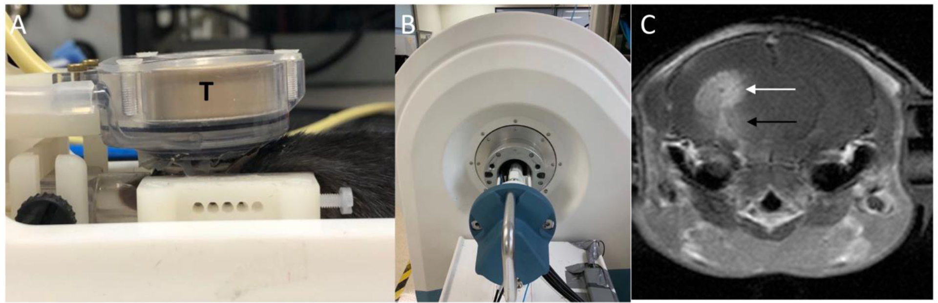
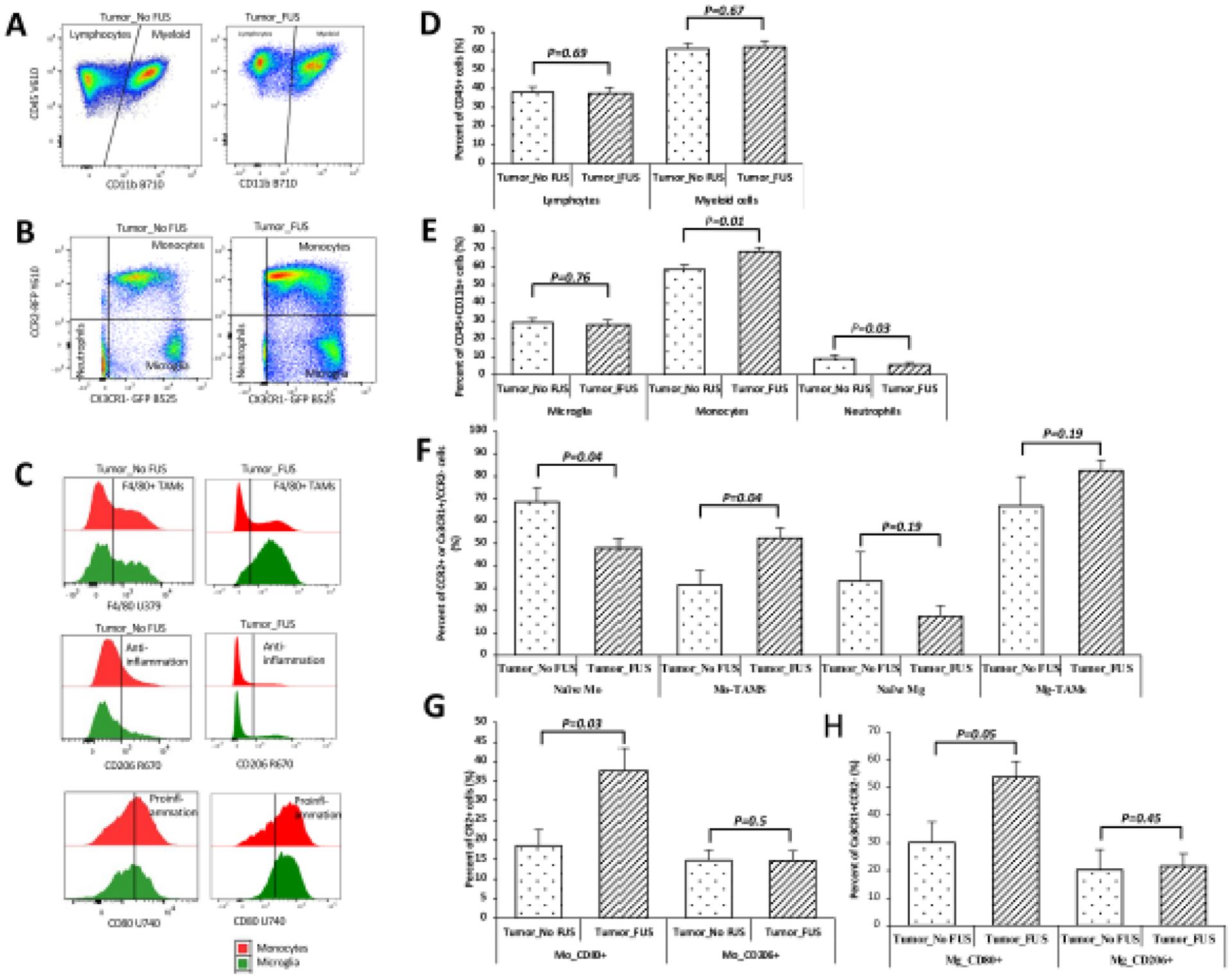
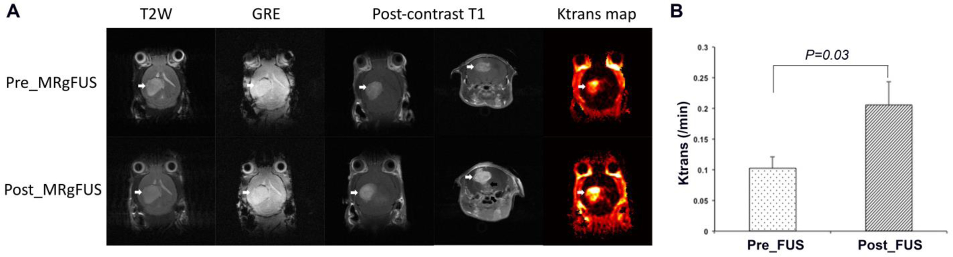
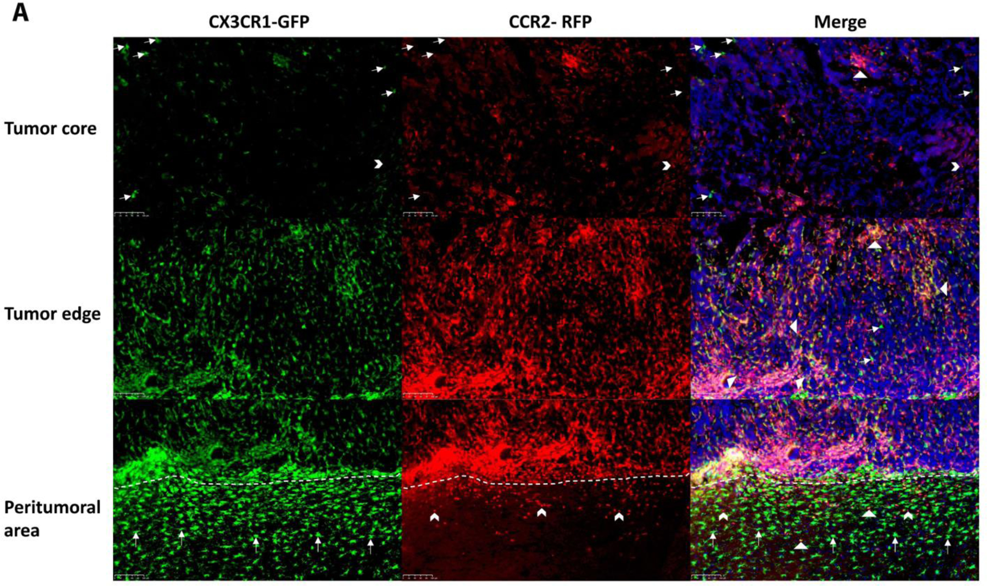
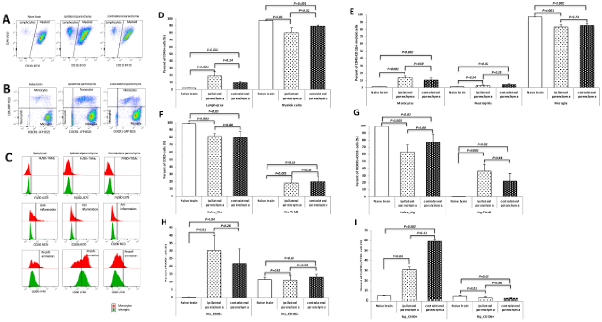
An orthotopically allografted mouse GL26 glioma model (Ccr2-Cx3cr1) was used to evaluate the effect of transient, focal opening of the blood-brain barrier (BBB) on the composition of tumor-associated macrophages and microglia (TAMs). BBB opening was induced by magnetic resonance imaging (MRI)-guided focused ultrasound (MRgFUS) combined with microbubbles. CX3CR1-GFP cells and CCR2-RFP cells in brain tumors were quantified in microscopic images. Tumors in animals treated with a single session of MRgFUS did not exhibit significant changes in cell numbers when compared with tumors in animals not receiving FUS. However, tumors that received two or three sessions of MRgFUS had significantly increased amounts of both CX3CR1-GFP and CCR2-RFP cells. The effect of MRgFUS on immune cell composition was also characterized and quantified using flow cytometry. Glioma implantation resulted in increased amounts of lymphocytes, monocytes and neutrophils in the brain parenchyma. Tumors administered MRgFUS exhibited increased numbers of monocytes and monocyte-derived TAMs. In addition, MRgFUS-treated tumors exhibited more CD80+ cells in monocytes and microglia. In summary, transient, focal opening of the BBB using MRgFUS combined with microbubbles can activate the homing and differentiation of monocytes and induce a shift toward a more pro-inflammatory status of the immune environment in glioblastoma.
我们使用原位同种异体移植 GL26 神经胶质瘤小鼠模型(Ccr2-Cx3cr1)来评估短暂、局部血脑屏障(BBB)开放对肿瘤相关巨噬细胞和小胶质细胞(TAMs)组成的影响。通过磁共振成像(MRI)引导的聚焦超声(MRgFUS)联合微泡来诱导 BBB 开放。在显微镜图像中定量分析脑肿瘤中的 CX3CR1-GFP 细胞和 CCR2-RFP 细胞。与未接受 FUS 的动物相比,单次 MRgFUS 治疗的动物的肿瘤中细胞数量没有明显变化。然而,接受两次或三次 MRgFUS 治疗的肿瘤中,CX3CR1-GFP 和 CCR2-RFP 细胞的数量明显增加。还使用流式细胞术对 MRgFUS 对免疫细胞组成的影响进行了特征描述和定量分析。脑实质中植入神经胶质瘤会导致淋巴细胞、单核细胞和中性粒细胞数量增加。接受 MRgFUS 治疗的肿瘤显示单核细胞和单核细胞衍生的 TAMs 数量增加。此外,MRgFUS 治疗的肿瘤中的单核细胞和小胶质细胞中 CD80+细胞增多。总之,使用 MRgFUS 联合微泡短暂、局部开放 BBB 可以激活单核细胞的归巢和分化,并诱导胶质母细胞瘤中免疫环境向更具炎症性状态的转变。