Department of Ophthalmology, Inselspital, Bern University Hospital, University of Bern, Bern, Switzerland.
Department of BioMedical Research, University of Bern, Bern, Switzerland.
J Neuroinflammation. 2023 Feb 4;20(1):25. doi: 10.1186/s12974-023-02712-1.
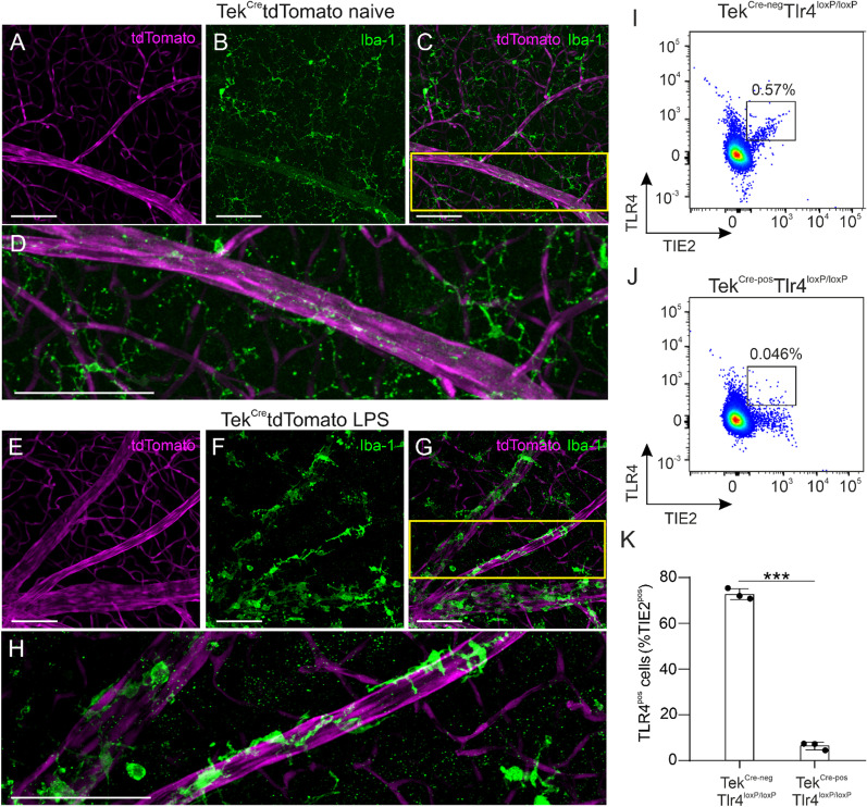
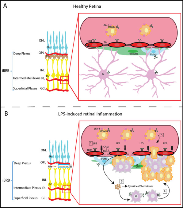
Clustering of microglia around the vasculature has been reported in the retina and the brain after systemic administration of lipopolysaccharides (LPS) in mice. LPS acts via activation of Toll-like receptor 4 (TRL4), which is expressed in several cell types including microglia, monocytes and vascular endothelial cells. The purpose of this study was to investigate the effect of systemic LPS in the pigmented mouse retina and the involvement of endothelial TLR4 in LPS-induced retinal microglia activation.
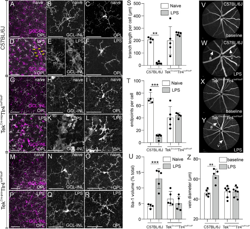
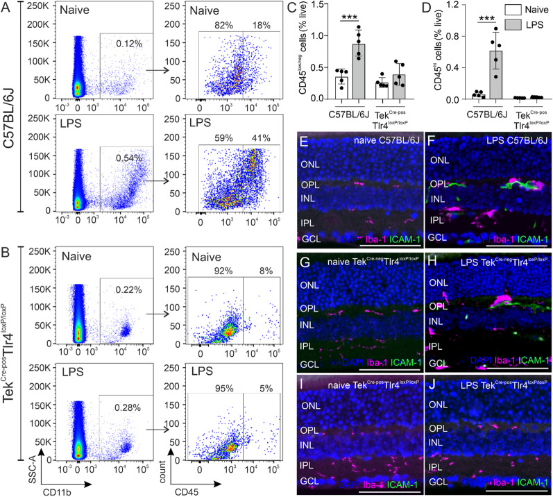
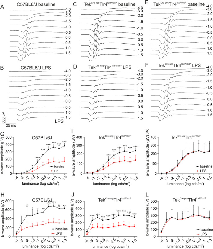
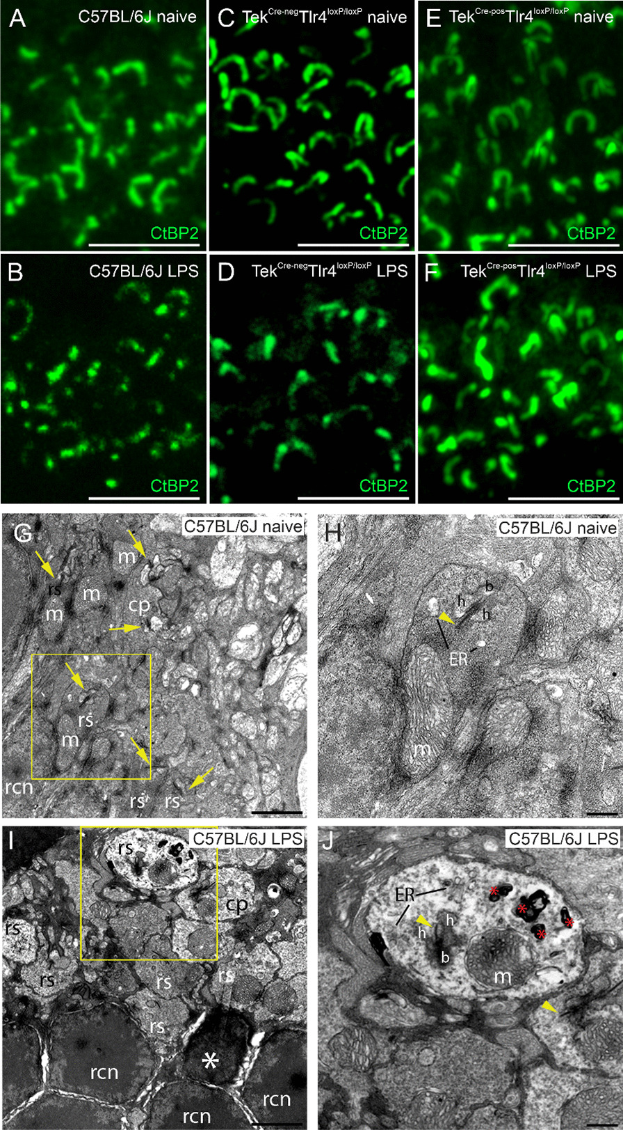
C57BL/6J, conditional knockout mice that lack Tlr4 expression selectively on endothelial cells (TekTlr4) and TekTlr4 mice were used. The mice were injected with 1 mg/kg LPS via the tail vein once per day for a total of 4 days. Prior to initiation of LPS injections and approximately 5 h after the last injection, in vivo imaging using fluorescein angiography and spectral-domain optical coherence tomography was performed. Immunohistochemistry, flow cytometry, electroretinography and transmission electron microscopy were utilized to investigate the role of endothelial TLR4 in LPS-induced microglia activation and retinal function.
Activation of microglia, infiltration of monocyte-derived macrophages, impaired ribbon synapse organization and retinal dysfunction were observed after the LPS exposure in C57BL/6J and TekTlr4 mice. None of these effects were observed in the retinas of conditional Tlr4 knockout mice after the LPS challenge.
The findings of the present study suggest that systemic LPS exposure can have detrimental effects in the healthy retina and that TLR4 expressed on endothelial cells is essential for retinal microglia activation and retinal dysfunction upon systemic LPS challenge. This important finding provides new insights into the role of microglia-endothelial cell interaction in inflammatory retinal disease.
在小鼠全身给予脂多糖 (LPS) 后,已报道在视网膜和大脑中血管周围存在小胶质细胞聚集。LPS 通过 Toll 样受体 4 (TLR4) 的激活起作用,TLR4 在包括小胶质细胞、单核细胞和血管内皮细胞在内的几种细胞类型中表达。本研究的目的是研究全身 LPS 在色素性小鼠视网膜中的作用以及内皮 TLR4 在 LPS 诱导的视网膜小胶质细胞激活中的作用。
使用 C57BL/6J、选择性缺乏内皮细胞 TLR4 表达的条件敲除小鼠 (TekTlr4) 和 TekTlr4 小鼠。将小鼠通过尾静脉每天注射 1 mg/kg LPS,共 4 天。在开始 LPS 注射之前和最后一次注射后约 5 小时,通过荧光素血管造影和光谱域光相干断层扫描进行体内成像。免疫组织化学、流式细胞术、视网膜电图和透射电子显微镜用于研究内皮 TLR4 在 LPS 诱导的小胶质细胞激活和视网膜功能中的作用。
在 C57BL/6J 和 TekTlr4 小鼠中,LPS 暴露后观察到小胶质细胞激活、单核细胞衍生巨噬细胞浸润、 ribbonsynapse 组织受损和视网膜功能障碍。在 LPS 挑战后,条件性 TLR4 敲除小鼠的视网膜中未观察到这些影响。
本研究的结果表明,全身 LPS 暴露会对健康的视网膜产生有害影响,并且内皮细胞表达的 TLR4 对于全身 LPS 挑战后视网膜小胶质细胞激活和视网膜功能障碍是必不可少的。这一重要发现为小胶质细胞-内皮细胞相互作用在炎症性视网膜疾病中的作用提供了新的见解。