Private Practice for Oral Surgery, Paradiesplatz 7-13, 88131, Lindau, Lake Constance, Germany.
Department of Oral and Maxillofacial Surgery, University Medical Center, Johannes Gutenberg University of Mainz, Augustusplatz 2, 55131, Mainz, Germany.
Int J Implant Dent. 2023 Feb 5;9(1):3. doi: 10.1186/s40729-023-00467-1.
This retrospective cohort study evaluates the regeneration of severe peri-implantitis deficiencies treated with the laser-assisted peri-implant defect regeneration (LAPIDER) approach within a 3-year follow-up.
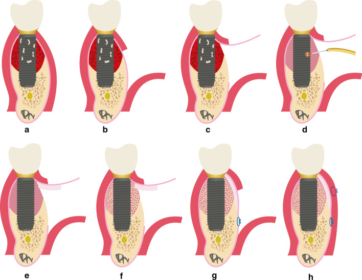
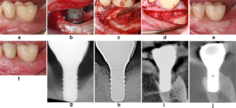
Twenty-four implants with severe peri-implantitis in 18 patients were treated according to the LAPIDER technique. In contrast to classic techniques for reconstructive peri-implantitis surgery with a marginal incision, a buccal split-flap preparation avoiding papillae separation was used. After a coronal flap elevation and a laser-assisted peri-implant defect cleaning, connective tissue and autogenous bone grafting was performed. Primary outcomes were the changes of the marginal bone levels (MBL) and the buccal bone thickness. Secondary outcomes included implant survival, peri-implant probing depths (PPD), bleeding on probing (BOP), recession, width of keratinized mucosa (KMW), thickness of keratinized mucosa (KMT), soft tissue esthetics (PES), and implant success.
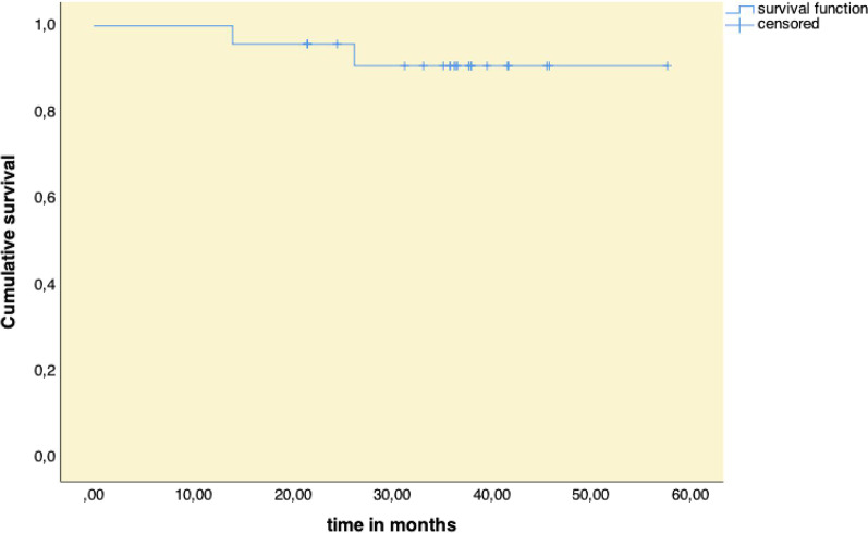
MBL improved interproximal by 3.10 ± 2.02 mm (p < 0.001), buccal by 3.49 ± 2.89 mm (p < 0.001), and lingual by 1.46 ± 1.98 mm (p = 0.003); buccal bone thickness by 0.55 ± 0.60 mm (p = 0.005), and 1.01 ± 1.25 mm (p = 0.001) at 1 and 3 mm below reference level. Two implants were removed; 22 implants were still in function at a mean follow-up of 36 months. PPD changed from 5.05 ± 1.39 to 3.08 ± 0.71 mm (p < 0.001); recession was reduced from 2.07 ± 1.70 to 0.91 ± 1.13 mm (p = 0.001); KMW increased from 2.91 ± 1.81 to 4.18 ± 1.67 mm (p = 0.006); KMT improved from 1.73 ± 0.50 to 2.44 ± 0.43 mm (p < 0.001); PES changed from 7.7 ± 2.8 to 10.7 ± 1.9 (p < 0.001). 45.8% to 54.2% of the implants met the criteria of implant success.
The favorable results document the proof of principle for the regeneration of severe peri-implant hard and soft tissue deficiencies by the LAPIDER treatment approach.
本回顾性队列研究评估了激光辅助种植体周围缺损再生(LAPIDER)方法治疗重度种植体周围炎缺损在 3 年随访期内的再生情况。
18 名患者的 24 个种植体患有严重的种植体周围炎,采用 LAPIDER 技术进行治疗。与经典的种植体周围炎重建手术的边缘切口技术不同,采用了避免乳头分离的颊侧瓣劈开准备。在进行冠向瓣提升和激光辅助种植体周围缺损清洁后,进行了结缔组织和自体骨移植。主要结果是边缘骨水平(MBL)和颊侧骨厚度的变化。次要结果包括种植体存活率、种植体探诊深度(PPD)、探诊出血(BOP)、退缩、角化黏膜宽度(KMW)、角化黏膜厚度(KMT)、软组织美学(PES)和种植体成功。
MBL 在近中方向改善了 3.10±2.02mm(p<0.001),颊侧方向改善了 3.49±2.89mm(p<0.001),舌侧方向改善了 1.46±1.98mm(p=0.003);颊侧骨厚度改善了 0.55±0.60mm(p=0.005)和 1.01±1.25mm(p=0.001),分别在参考水平以下 1mm 和 3mm 处。有 2 个种植体被取出;22 个种植体在平均 36 个月的随访中仍在使用。PPD 从 5.05±1.39mm 降至 3.08±0.71mm(p<0.001);退缩从 2.07±1.70mm 降至 0.91±1.13mm(p=0.001);KMW 从 2.91±1.81mm 增加至 4.18±1.67mm(p=0.006);KMT 从 1.73±0.50mm 增加至 2.44±0.43mm(p<0.001);PES 从 7.7±2.8 改善至 10.7±1.9(p<0.001)。45.8%至 54.2%的种植体符合种植体成功的标准。
这些良好的结果证明了 LAPIDER 治疗方法在重度种植体周围软硬组织缺损再生方面的原理证明。