Rana Deepti, Padmanaban Prasanna, Becker Malin, Stein Fabian, Leijten Jeroen, Koopman Bart, Rouwkema Jeroen
Department of Biomechanical Engineering, Technical Medical Centre, Faculty of Engineering Technology, University of Twente, 7522NB Enschede, the Netherlands.
Department of Developmental BioEngineering, Faculty of Science and Technology, Technical Medical Centre, University of Twente, 7522NB Enschede, the Netherlands.
Mater Today Bio. 2023 Jan 20;19:100551. doi: 10.1016/j.mtbio.2023.100551. eCollection 2023 Apr.
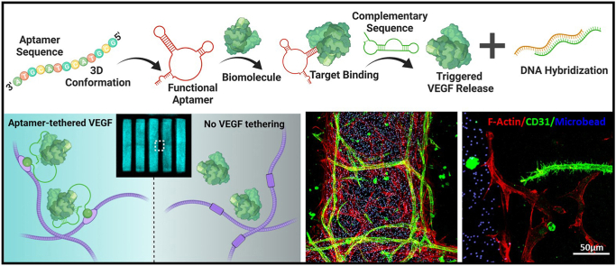
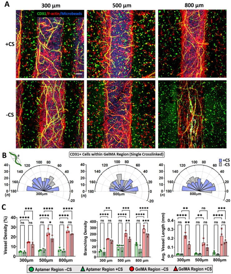
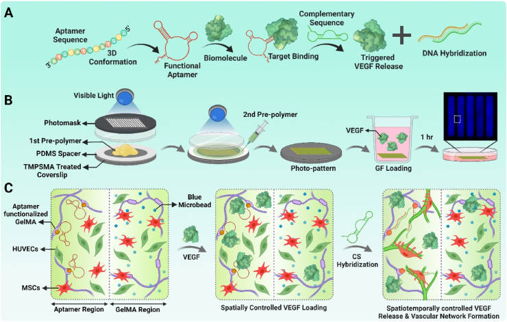
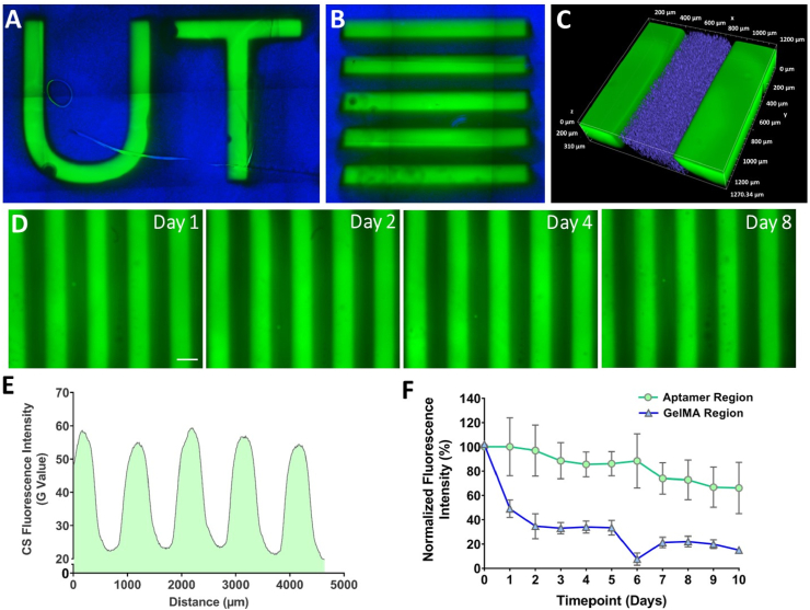
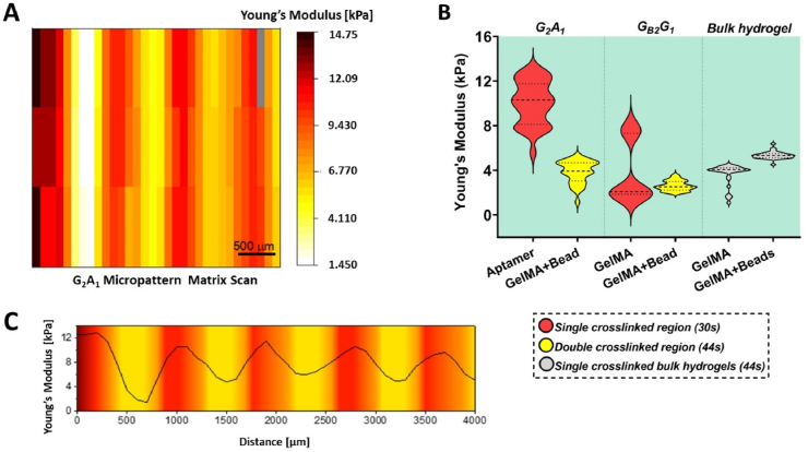
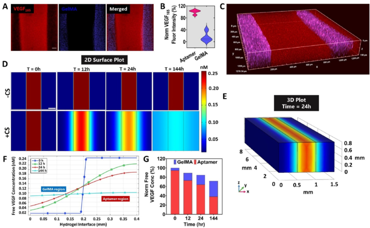
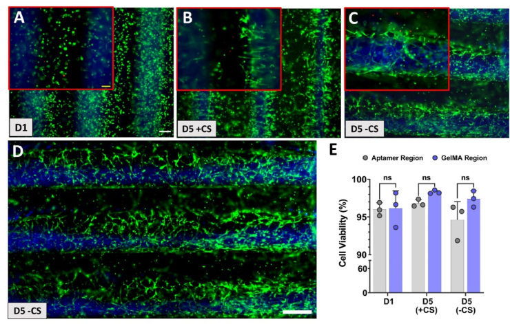
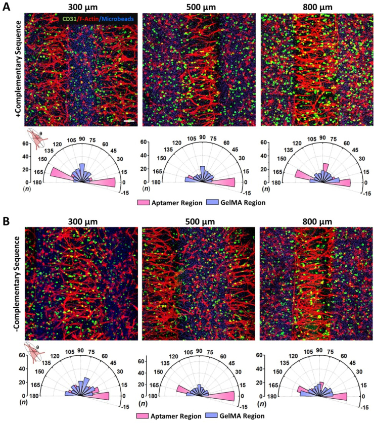
Given the dynamic nature of engineered vascular networks within biofabricated tissue analogues, it is instrumental to have control over the constantly evolving biochemical cues within synthetic matrices throughout tissue remodeling. Incorporation of pro-angiogenic vascular endothelial growth factor (VEGF) specific aptamers into cell-instructive polymer networks is shown to be pivotal for spatiotemporally controlling the local bioactivity of VEGF that selectively elicit specific cell responses. To harness this effect and quantitatively unravel its spatial resolution, herein, bicomponent micropatterns consisting of VEGF specific aptamer-functionalized gelatin methacryloyl (GelMA) (aptamer regions) overlaid with pristine GelMA regions using visible-light photoinitiators (Ru/SPS) were fabricated via two-step photopatterning approach. For the 3D co-culture study, human umbilical vein-derived endothelial cells and mesenchymal stromal cells were used as model cell types. Bicomponent micropatterns with spatially defined spacings (300/500/800 μm) displayed high aptamer retention, aptamer-fluorescent complementary sequence (CS) molecular recognition and VEGF sequestration localized within patterned aptamer regions. Stiffness gradient at the interface of aptamer and GelMA regions was observed with high modulus in aptamer region followed by low stiffness GelMA regions. Leveraging aptamer-tethered VEGF's dynamic affinity interactions with CS that upon hybridization facilitates triggered VEGF release, co-culture studies revealed unique characteristics of aptamer-tethered VEGF to form spatially defined luminal vascular networks covered with filopodia-like structures (spatial control) and highlights their ability to control network properties including orientation over time using CS as an external trigger (temporal control). Moreover, the comparison of single and double exposed regions within micropatterns revealed differences in cell behavior among both regions. Specifically, the localized aptamer-tethered VEGF within single exposed aptamer regions exhibited higher cellular alignment within the micropatterns till d5 of culture. Taken together, this study highlights the potential of photopatterned aptamer-tethered VEGF to spatiotemporally regulate vascular morphogenesis as a tool for controlling vascular remodeling
鉴于生物制造的组织类似物中工程化血管网络的动态性质,在整个组织重塑过程中控制合成基质内不断演变的生化信号是很有帮助的。将促血管生成的血管内皮生长因子(VEGF)特异性适配体整合到具有细胞指导作用的聚合物网络中,对于时空控制VEGF的局部生物活性至关重要,VEGF的局部生物活性可选择性地引发特定的细胞反应。为了利用这种效应并定量揭示其空间分辨率,本文通过两步光图案化方法制备了双组分微图案,该微图案由VEGF特异性适配体功能化的甲基丙烯酸明胶(GelMA)(适配体区域)和使用可见光光引发剂(Ru/SPS)覆盖的原始GelMA区域组成。对于三维共培养研究,使用人脐静脉来源的内皮细胞和间充质基质细胞作为模型细胞类型。具有空间定义间距(300/500/800μm)的双组分微图案显示出高适配体保留率、适配体-荧光互补序列(CS)分子识别以及VEGF隔离定位在图案化的适配体区域内。在适配体和GelMA区域的界面处观察到刚度梯度,适配体区域具有高模量,随后是低刚度的GelMA区域。利用适配体连接的VEGF与CS的动态亲和相互作用,杂交后促进VEGF释放,共培养研究揭示了适配体连接的VEGF形成由丝状伪足样结构覆盖的空间定义的管腔血管网络的独特特征(空间控制),并突出了它们使用CS作为外部触发因素随时间控制包括取向在内的网络特性的能力(时间控制)。此外,微图案内单次和两次曝光区域的比较揭示了两个区域之间细胞行为的差异。具体而言,单次曝光适配体区域内局部化的适配体连接的VEGF在培养至第5天时在微图案内表现出更高的细胞排列。综上所述,本研究强调了光图案化的适配体连接的VEGF作为控制血管重塑的工具在时空调节血管形态发生方面的潜力