• 文献检索
  • 文档翻译
  • 深度研究
  • 学术资讯
  • Suppr Zotero 插件Zotero 插件
  • 邀请有礼
  • 套餐&价格
  • 历史记录
应用&插件
Suppr Zotero 插件Zotero 插件浏览器插件Mac 客户端Windows 客户端微信小程序
定价
高级版会员购买积分包购买API积分包
服务
文献检索文档翻译深度研究API 文档MCP 服务
关于我们
关于 Suppr公司介绍联系我们用户协议隐私条款
关注我们

Suppr 超能文献

核心技术专利:CN118964589B侵权必究
粤ICP备2023148730 号-1Suppr @ 2026

文献检索

告别复杂PubMed语法,用中文像聊天一样搜索,搜遍4000万医学文献。AI智能推荐,让科研检索更轻松。

立即免费搜索

文件翻译

保留排版,准确专业,支持PDF/Word/PPT等文件格式,支持 12+语言互译。

免费翻译文档

深度研究

AI帮你快速写综述,25分钟生成高质量综述,智能提取关键信息,辅助科研写作。

立即免费体验

500例液基薄层巴氏涂片的BestCyte®初步筛查:3位细胞病理学家之间的观察者间诊断一致性,与相对于真实参考诊断的谓词手工显微镜检查结果相比,确定了无上皮内病变或恶性病变(NILM)、意义不明确的非典型鳞状细胞(ASCUS+)、低度鳞状上皮内病变(LSIL+)和非典型鳞状细胞不能排除高级别鳞状上皮内病变(ASCH+)阈值的特异性、敏感性和等效性分级。

BestCyte® primary screening of 500 ThinPrep Pap Test thin-layers: 3 Cytologists' Interobserver diagnostic concordance with predicate manual microscopy relative to Truth Reference diagnoses defining NILM, ASCUS+, LSIL+, and ASCH+ thresholds for specificity, sensitivity, and equivalency grading.

作者信息

Chantziantoniou Nikolaos

机构信息

CellPathology Plus, 136 Silver Linden Drive, Richmond Hill, Ontario L4B4H6, Canada.

出版信息

J Pathol Inform. 2023 Jan 2;14:100182. doi: 10.1016/j.jpi.2022.100182. eCollection 2023.

DOI:10.1016/j.jpi.2022.100182
PMID:36747889
原文链接:https://pmc.ncbi.nlm.nih.gov/articles/PMC9898738/
Abstract

BACKGROUND

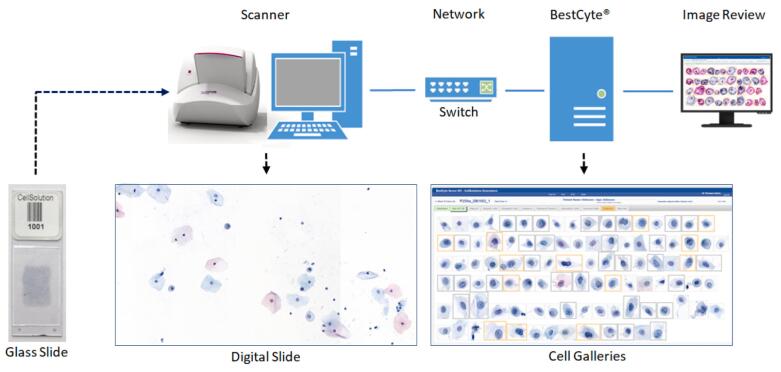
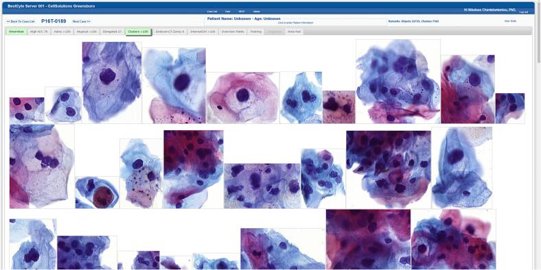
The BestCyte® Cell Sorter Imaging System (BestCyte) facilitates algorithmic discrimination of clinically relevant cells in Pap test cytopathology by classifying and projecting images of cells in galleries based on cytomorphology. Warranted is awareness of potential BestCyte advantages as measured through 3 cytologists' interobserver diagnostic concordance, specificity and sensitivity differentials, and equivalency grading relative to manual microscopy (MM).

OBJECTIVES

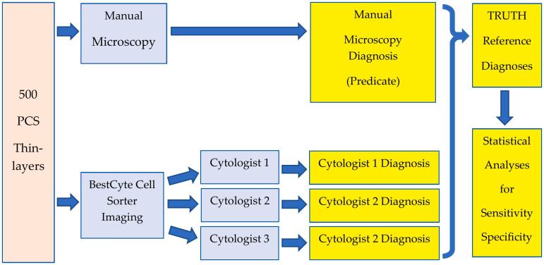
Using 500 MM-reported ThinPrep thin-layers, analyze: (1) cytologists' blinded BestCyte screening to raise Bethesda diagnoses; (2) correlate BestCyte and MM diagnoses (i.e., predicate) to establish Truth Reference Diagnoses (TRDx) from concordance between 4 possible diagnoses; (3) analyze cytologists' and MM predicate diagnoses through 4 diagnostic thresholds defined by TRDx: NILM () for specificity, and ASCUS+, LSIL+, and ASCH+ () for graded sensitivity (with abnormal cells decreasing in size with increasing dysplasia); and, (4) statistically determine cytologists' equivalency grading to MM using 95% Confidence Interval (CI) ranges.

RESULTS

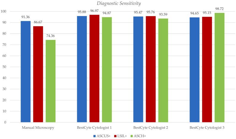
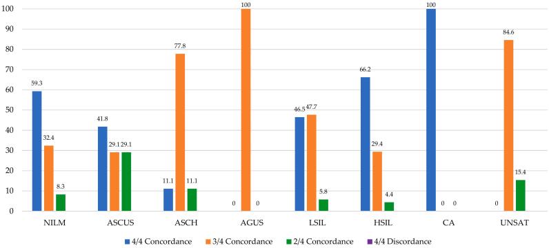
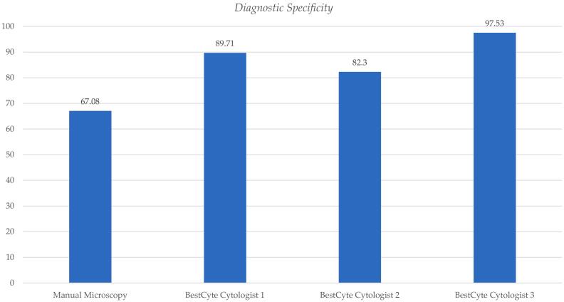
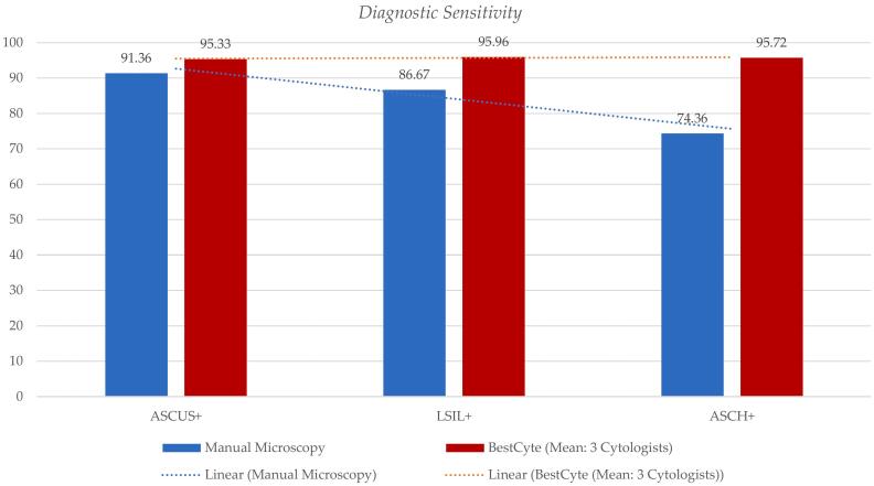
500 TRDx breakdown (n/%): NILM (241/48.2), ASCUS (79/15.8), ASCH (9/1.80), AGUS (2/0.40), LSIL (86/17.2), HSIL (68/13.6), CA (2/0.40), UNSAT (13/2.60). TRDx breakdown (n/%) per 4 of 4, 3 of 4, 2 of 4 diagnostic concordances: 264 (52.8%), 182 (36.4%), 54 (10.8%), respectively. No cases of discordant diagnoses were recorded. HSIL TRDx were established from 66.2% of 4 of 4 concordances, followed by NILM (59.3%), LSIL (46.5%), ASCUS (41.8%); antithetically, from 4.40% of 2 of 4 concordances. Specificity for MM predicate (NILM): 67.08%; for Cytologists 1, 2, and 3: 89.71%, 82.30%, 97.53%, respectively. For NILM threshold, cytologists revealed equivalency to MM. Sensitivity for ASCUS+, LSIL+, and ASCH+ thresholds: MM (91.36%, 86.67%, 74.36%); Cytologist 1 (95.88%, 96.97%, 94.87%); Cytologist 2 (95.47%, 95.76%, 93.59%), Cytologist 3 (94.65%, 95.15%, 98.72%), respectively. Cytologists revealed equivalency to MM for graded thresholds; with Cytologist 3 for ASCUS+ being: .

CONCLUSIONS

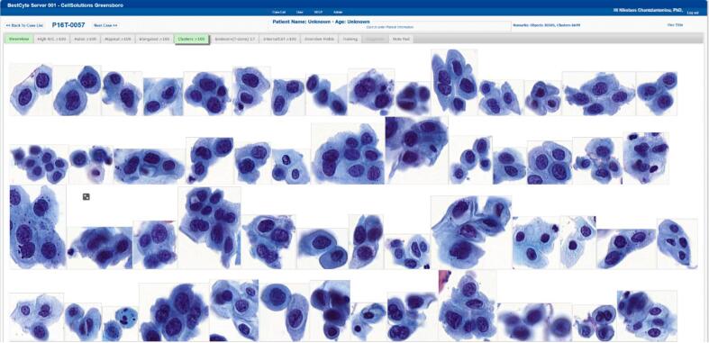
BestCyte detects and efficiently displays abnormal cells in strategic galleries standardizing objectivity by systematizing mosaics of cell-types for cytologists' consideration. BestCyte fosters consistent, enhanced cytologists' sensitivity values for the ASCUS+, LSIL+, and ASCH+ thresholds relative to MM. Also, BestCyte facilitates improved specificity and superior equivalency grading to MM reflecting efficient screening, and reduced labor. Confident interpretations of small dysplastic epithelial cells characteristic of HSIL led to exceptional interobserver diagnostic concordance inferring BestCyte is primed for effective cervical cancer screening practice.

摘要

背景

BestCyte®细胞分选成像系统(BestCyte)通过根据细胞形态对图库中的细胞图像进行分类和投影,有助于在巴氏试验细胞病理学中对临床相关细胞进行算法鉴别。需要了解通过3位细胞病理学家的观察者间诊断一致性、特异性和敏感性差异以及相对于手动显微镜检查(MM)的等效性分级所衡量的BestCyte的潜在优势。

目的

使用500份MM报告的ThinPrep薄层涂片,分析:(1)细胞病理学家对BestCyte进行盲法筛查以提高贝塞斯达诊断;(2)关联BestCyte和MM诊断(即预测),以便根据4种可能诊断之间的一致性建立真实参考诊断(TRDx);(3)通过TRDx定义的4个诊断阈值分析细胞病理学家和MM的预测诊断:NILM()用于特异性,ASCUS+、LSIL+和ASCH+()用于分级敏感性(异常细胞随着发育异常程度增加而变小);以及(4)使用95%置信区间(CI)范围通过统计学方法确定细胞病理学家相对于MM的等效性分级。

结果

500份TRDx分类(n/%):NILM(241/48.2),ASCUS(79/15.8),ASCH(9/1.80),AGUS(2/0.40),LSIL(86/17.2),HSIL(68/13.6),CA(2/0.40),UNSAT(13/2.60)。每4种诊断一致性中的TRDx分类(n/%):4种诊断全一致的为264(52.8%),3种诊断一致的为182(36.4%),2种诊断一致的为54(10.8%)。未记录到诊断不一致的病例。HSIL的TRDx由4种诊断全一致的病例中的66.2%确定,其次是NILM(59.3%)、LSIL(46.5%)、ASCUS(41.8%);相反,由2种诊断一致的病例中的4.40%确定。MM预测(NILM)的特异性:67.08%;细胞病理学家1、2和3的特异性分别为89.71%、82.30%、97.53%。对于NILM阈值,细胞病理学家显示与MM等效。ASCUS+、LSIL+和ASCH+阈值的敏感性:MM(91.36%、86.67%、74.36%);细胞病理学家1(95.88%)、96(96.97%)、94.87%);细胞病理学家2(95.47%、95.76%、93.59%),细胞病理学家3(94.65%、95.15%、98.72%)。细胞病理学家显示在分级阈值方面与MM等效;对于ASCUS+,细胞病理学家三为:。

结论

BestCyte通过将细胞类型的镶嵌图系统化以供细胞病理学家考虑,在策略性图库中检测并有效显示异常细胞,从而实现客观性标准化。相对于MM,BestCyte提高了细胞病理学家对ASCUS+、LSIL+和ASCH+阈值的敏感性值,且具有一致性。此外,BestCyte有助于提高特异性,并在等效性分级方面优于MM,体现了高效筛查和减少劳动量。对HSIL特有的小发育异常上皮细胞的可靠解读导致了观察者间出色的诊断一致性,这表明BestCyte适合用于有效的宫颈癌筛查实践。

https://cdn.ncbi.nlm.nih.gov/pmc/blobs/7de3/9898738/c8ffc89c8de2/gr8.jpg
https://cdn.ncbi.nlm.nih.gov/pmc/blobs/7de3/9898738/b9a7325e5659/gr1.jpg
https://cdn.ncbi.nlm.nih.gov/pmc/blobs/7de3/9898738/368944d7f30f/gr2.jpg
https://cdn.ncbi.nlm.nih.gov/pmc/blobs/7de3/9898738/03cd5bb1fd70/gr3.jpg
https://cdn.ncbi.nlm.nih.gov/pmc/blobs/7de3/9898738/8292ad28f9ad/gr4.jpg
https://cdn.ncbi.nlm.nih.gov/pmc/blobs/7de3/9898738/206e8c82482a/gr5.jpg
https://cdn.ncbi.nlm.nih.gov/pmc/blobs/7de3/9898738/2d4d32dacf8d/gr6.jpg
https://cdn.ncbi.nlm.nih.gov/pmc/blobs/7de3/9898738/915c6be45708/gr7.jpg
https://cdn.ncbi.nlm.nih.gov/pmc/blobs/7de3/9898738/c8ffc89c8de2/gr8.jpg

相似文献

1
BestCyte® primary screening of 500 ThinPrep Pap Test thin-layers: 3 Cytologists' Interobserver diagnostic concordance with predicate manual microscopy relative to Truth Reference diagnoses defining NILM, ASCUS+, LSIL+, and ASCH+ thresholds for specificity, sensitivity, and equivalency grading.500例液基薄层巴氏涂片的BestCyte®初步筛查:3位细胞病理学家之间的观察者间诊断一致性,与相对于真实参考诊断的谓词手工显微镜检查结果相比,确定了无上皮内病变或恶性病变(NILM)、意义不明确的非典型鳞状细胞(ASCUS+)、低度鳞状上皮内病变(LSIL+)和非典型鳞状细胞不能排除高级别鳞状上皮内病变(ASCH+)阈值的特异性、敏感性和等效性分级。
J Pathol Inform. 2023 Jan 2;14:100182. doi: 10.1016/j.jpi.2022.100182. eCollection 2023.
2
BestCyte® Cell Sorter Imaging System: Primary and adjudicative whole slide image rescreening review times of 500 ThinPrep Pap test thin-layers - An intra-observer, time-surrogate analysis of diagnostic confidence potentialities.BestCyte®细胞分选成像系统:500例ThinPrep巴氏试验薄层的初次和判定性全玻片图像重新筛查复查时间——诊断置信度潜力的观察者内、时间替代分析
J Pathol Inform. 2022 May 21;13:100095. doi: 10.1016/j.jpi.2022.100095. eCollection 2022.
3
Evaluation of CellSolutions BestPrep® automated thin-layer liquid-based cytology Papanicolaou slide preparation and BestCyte® cell sorter imaging system.CellSolutions BestPrep® 自动薄层液基细胞学巴氏涂片制备及 BestCyte® 细胞分选成像系统的评估
Acta Cytol. 2014;58(5):469-77. doi: 10.1159/000367837. Epub 2014 Sep 27.
4
[Health technology assessment report: Computer-assisted Pap test for cervical cancer screening].[卫生技术评估报告:用于宫颈癌筛查的计算机辅助巴氏试验]
Epidemiol Prev. 2012 Sep-Oct;36(5 Suppl 3):e1-43.
5
Diagnostic accuracy of oral cancer cytology in a pilot study.一项初步研究中口腔癌细胞学检查的诊断准确性
Diagn Pathol. 2017 Mar 16;12(1):27. doi: 10.1186/s13000-017-0618-3.
6
Role of Pap Test terminology and age in the detection of carcinoma invasive and carcinoma in situ in medically underserved California women.巴氏试验术语和年龄在加利福尼亚州医疗服务不足的女性浸润性癌和原位癌检测中的作用。
Diagn Cytopathol. 2004 Apr;30(4):227-34. doi: 10.1002/dc.10306.
7
Resolving ASCUS without recourse to HPV testing: manual reprocessing of residual automated liquid-based cytology (ALBC) material using manual liquid-based cytology (MLBC).不借助HPV检测解决非典型鳞状细胞意义不明确(ASCUS)的问题:使用手工液基细胞学(MLBC)对剩余的自动化液基细胞学(ALBC)材料进行手工再处理。
Diagn Cytopathol. 2005 Dec;33(6):434-40. doi: 10.1002/dc.20324.
8
Cytology versus HPV testing for cervical cancer screening in the general population.普通人群宫颈癌筛查中细胞学检查与HPV检测的比较
Cochrane Database Syst Rev. 2017 Aug 10;8(8):CD008587. doi: 10.1002/14651858.CD008587.pub2.
9
Assisted primary screening using the automated ThinPrep Imaging System.使用自动薄层液基细胞学成像系统进行辅助初次筛查。
Am J Clin Pathol. 2005 Feb;123(2):281-7.
10
Triage of women with ASCUS and LSIL on Pap smear reports: management by repeat Pap smear, HPV DNA testing, or colposcopy?巴氏涂片报告中不典型鳞状细胞(ASCUS)和低度鳞状上皮内病变(LSIL)女性的分流:通过重复巴氏涂片、人乳头瘤病毒(HPV)DNA检测或阴道镜检查进行管理?
J Fam Pract. 1998 Feb;46(2):125-34.

引用本文的文献

1
Assessment of the efficacy and accuracy of cervical cytology screening with the Hologic Genius Digital Diagnostics System.使用Hologic Genius数字诊断系统评估宫颈细胞学筛查的有效性和准确性。
Cancer Cytopathol. 2025 Jul;133(7):e70022. doi: 10.1002/cncy.70022.
2
Performance of artificial intelligence for diagnosing cervical intraepithelial neoplasia and cervical cancer: a systematic review and meta-analysis.人工智能诊断宫颈上皮内瘤变和宫颈癌的性能:系统评价与荟萃分析
EClinicalMedicine. 2024 Dec 28;80:102992. doi: 10.1016/j.eclinm.2024.102992. eCollection 2025 Feb.
3
Artificial intelligence strengthenes cervical cancer screening - present and future.

本文引用的文献

1
BestCyte® Cell Sorter Imaging System: Primary and adjudicative whole slide image rescreening review times of 500 ThinPrep Pap test thin-layers - An intra-observer, time-surrogate analysis of diagnostic confidence potentialities.BestCyte®细胞分选成像系统:500例ThinPrep巴氏试验薄层的初次和判定性全玻片图像重新筛查复查时间——诊断置信度潜力的观察者内、时间替代分析
J Pathol Inform. 2022 May 21;13:100095. doi: 10.1016/j.jpi.2022.100095. eCollection 2022.
2
Relevance of the College of American Pathologists guideline for validating whole slide imaging for diagnostic purposes to cytopathology.美国病理学家学院关于验证全切片成像在诊断目的方面用于细胞病理学的指南的相关性。
Cytopathology. 2023 Jan;34(1):5-14. doi: 10.1111/cyt.13178. Epub 2022 Sep 26.
3
人工智能增强宫颈癌筛查 - 现状与未来。
Cancer Biol Med. 2024 Sep 19;21(10):864-79. doi: 10.20892/j.issn.2095-3941.2024.0198.
4
Artificial Intelligence-driven Digital Cytology-based Cervical Cancer Screening: Is the Time Ripe to Adopt This Disruptive Technology in Resource-constrained Settings? A Literature Review.人工智能驱动的数字细胞学宫颈癌筛查:在资源有限的环境中采用这种颠覆性技术是否时机成熟?文献综述。
J Digit Imaging. 2023 Aug;36(4):1643-1652. doi: 10.1007/s10278-023-00821-0. Epub 2023 Apr 7.
Rethinking Algorithm Performance Metrics for Artificial Intelligence in Diagnostic Medicine.重新思考诊断医学中人工智能的算法性能指标
JAMA. 2022 Jul 26;328(4):329-330. doi: 10.1001/jama.2022.10561.
4
Understanding, explaining, and utilizing medical artificial intelligence.理解、解释和利用医学人工智能。
Nat Hum Behav. 2021 Dec;5(12):1636-1642. doi: 10.1038/s41562-021-01146-0. Epub 2021 Jun 28.
5
Experience Reviewing Digital Pap Tests using a Gallery of Images.使用图像库回顾数字巴氏涂片检查的经验
J Pathol Inform. 2021 Feb 23;12:7. doi: 10.4103/jpi.jpi_96_20. eCollection 2021.
6
Validating Whole Slide Imaging Systems for Diagnostic Purposes in Pathology.用于病理学诊断目的的全切片成像系统验证。
Arch Pathol Lab Med. 2022 Apr 1;146(4):440-450. doi: 10.5858/arpa.2020-0723-CP.
7
Artificial intelligence and computational pathology.人工智能与计算病理学。
Lab Invest. 2021 Apr;101(4):412-422. doi: 10.1038/s41374-020-00514-0. Epub 2021 Jan 16.
8
Computational Cytology: Lessons Learned from Pap Test Computer-Assisted Screening.计算细胞学:巴氏涂片计算机辅助筛查的经验教训。
Acta Cytol. 2021;65(4):286-300. doi: 10.1159/000508629. Epub 2020 Jul 21.
9
Comparison of ThinPrep Integrated Imager-Assisted Screening versus Manual Screening of ThinPrep Liquid-Based Cytology Specimens.ThinPrep集成成像仪辅助筛查与ThinPrep液基细胞学标本手工筛查的比较。
Acta Cytol. 2020;64(5):486-491. doi: 10.1159/000507910. Epub 2020 Jun 12.
10
EpithNet: Deep Regression for Epithelium Segmentation in Cervical Histology Images.EpithNet:用于宫颈组织学图像上皮细胞分割的深度回归
J Pathol Inform. 2020 Mar 30;11:10. doi: 10.4103/jpi.jpi_53_19. eCollection 2020.