Department of Orthopedics and Traumatology, Çekirge State Hospital, Bursa-Türkiye.
Department of Orthopedics and Traumatology, Erciyes University Faculty of Medicine, Kayseri-Türkiye.
Ulus Travma Acil Cerrahi Derg. 2023 Feb;29(2):247-251. doi: 10.14744/tjtes.2022.15163.
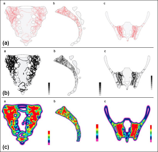
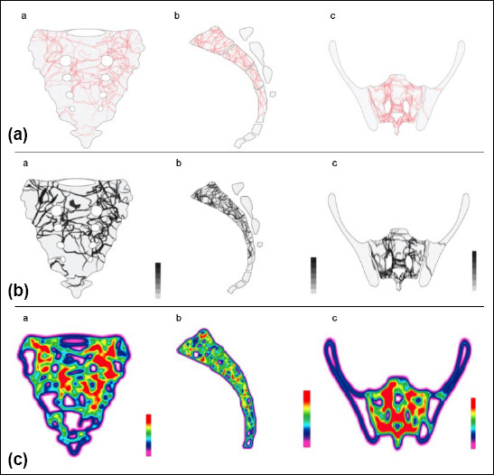
Sacral fractures are uncommon and understanding three-dimensional morphology is needed to obtain proper treatment. The purpose of this study was to identify the repeatable fracture patterns and comminution zones for traumatic sacral fractures and create fracture maps.
Computerized tomography images of 72 patients with traumatic sacral fracture were included in the study. For each fracture, fracture lines were identified and digitally reduced. All fractures were superimposed over a template and fracture maps; comminution zones and heatmaps were created for each zone.
There were 40 males and 32 females with a mean age of 46.5±19.9. Fifty-three (73.6%) patients sustained major trauma, and 19 (26.4%) had minor trauma. There were 37 (51.4%) Zone 1, 22 (30.6%) Zone 2, and 13 (18.1%) Zone 3 fractures. Each Denis zone showed certain fracture patterns. In Zone 1 fractures, most of the fracture lines were vertical and oblique (up to 45°) orientation on both sides. In Zone 2 fractures, fracture lines were concentrated on the S1 and S2 levels. Anterolateral and posterolateral parts of the sacrum were less affected in right-side fractures. In Zone 3 fractures, fractures were concentrated in S1, S2, and S3 levels around the sacral canal. The median sacral crest and midline remained mostly unaffected.
Sacral fractures showed specific repeatable patterns for each zone. These findings may be helpful for pre-operative planning, placement of fixation material, design of new implants, and modification of current fracture-classification systems.
骶骨骨折并不常见,为了获得恰当的治疗,了解其三维形态是必要的。本研究的目的是确定创伤性骶骨骨折的可重复骨折模式和粉碎区域,并创建骨折图谱。
本研究纳入了 72 例创伤性骶骨骨折患者的计算机断层扫描图像。对于每个骨折,识别骨折线并进行数字复位。所有骨折均与模板和骨折图谱叠加,为每个区域创建粉碎区和热点图。
男性 40 例,女性 32 例,平均年龄 46.5±19.9 岁。53 例(73.6%)患者遭受重大创伤,19 例(26.4%)患者遭受轻微创伤。Zone 1 骨折 37 例(51.4%),Zone 2 骨折 22 例(30.6%),Zone 3 骨折 13 例(18.1%)。每个 Denis 区都有特定的骨折模式。在 Zone 1 骨折中,大多数骨折线在两侧呈垂直和斜向(达 45°)方向。在 Zone 2 骨折中,骨折线集中在 S1 和 S2 水平。右侧骨折时,骶骨前外侧和后外侧部分受影响较小。在 Zone 3 骨折中,骨折集中在骶骨管周围的 S1、S2 和 S3 水平。骶骨中线和中嵴大多未受影响。
骶骨骨折在每个区域都表现出特定的可重复模式。这些发现可能有助于术前规划、固定材料的放置、新型植入物的设计以及对当前骨折分类系统的修改。