Faculty of Engineering, University of Toyama, 3190 Gofuku, Toyama, 93008555, Japan.
School of Medicine, Kanazawa Medical University, 1-1 Daigaku, Uchinada, Kahoku, Ishikawa, 9200293, Japan.
J Med Ultrason (2001). 2023 Apr;50(2):131-141. doi: 10.1007/s10396-023-01289-9. Epub 2023 Feb 9.
The contrasts of flowing blood in in vitro experiments using porcine blood and in vivo measurements of human jugular veins were analyzed to demonstrate that the hemorheological property was dependent on the shear rate.
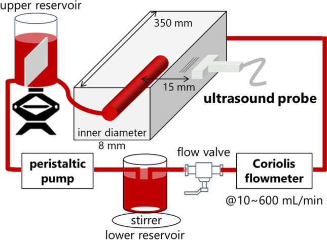
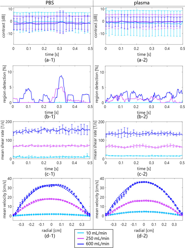
Blood samples (45% hematocrit) suspended in saline or plasma were compared with examine the difference in viscoelasticity. Ultrafast plane-wave imaging at an ultrasonic center frequency of 7.5 MHz was performed on different steady flows in a graphite-agar phantom. Also, in vivo measurement was performed in young, healthy subjects and patients with diabetes. A spatiotemporal matrix of beamformed radio-frequency data was used for the singular value decomposition (SVD) clutter filter. The clutter-filtered B-mode image was calculated as the amplitude envelope normalized at the first frame in the diastolic phase to evaluate contrast. The shear rate was estimated as the velocity gradient perpendicular to the lateral axis.
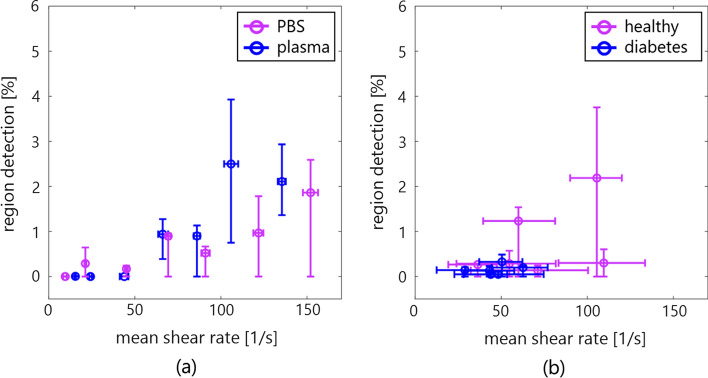
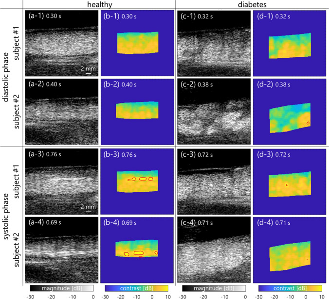
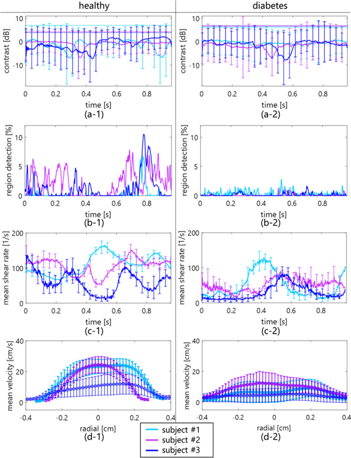
Although nonaggregated erythrocytes at a high shear rate exhibited a low echogenicity, the echogenicity in the plasma sample overall increased due to erythrocyte aggregation at a low shear rate. In addition, the frequency of detection of specular components, defined as components beyond twice the standard deviation of a contrast map obtained from a clutter-filtered B-mode image, increased in the porcine blood at a high shear rate and the venous blood in healthy subjects versus patients with diabetes.
The possibility of characterizing hemorheological properties dependent on the shear rate and diabetes condition was indicated using ultrafast plane-wave imaging with an SVD-based clutter filter.
分析在猪血液的体外实验和人类颈静脉的体内测量中血流对比,以证明血液流变学性质依赖于剪切率。
比较悬浮在盐水中或血浆中的血液样本(45%的血细胞比容),以检查粘弹性的差异。在石墨-琼脂模型中的不同稳定流中,使用超快速平面波成像以 7.5 MHz 的超声中心频率进行。此外,还在年轻、健康的受试者和糖尿病患者中进行了体内测量。使用波束形成射频数据的时空矩阵进行奇异值分解(SVD)杂波滤波器。杂波滤波后的 B 模式图像作为在舒张期第一帧处归一化的幅度包络进行计算,以评估对比度。剪切率估计为垂直于横向轴的速度梯度。
尽管在高剪切率下非聚集的红细胞表现出低回声性,但由于在低剪切率下红细胞聚集,整体上血浆样本的回声性增加。此外,在高剪切率下的猪血液和健康受试者的静脉血液中,定义为超出从杂波滤波的 B 模式图像获得的对比度图的两倍标准偏差的镜面反射成分的检测频率增加,而在糖尿病患者中则减少。
使用基于 SVD 的杂波滤波器的超快速平面波成像,表明了依赖于剪切率和糖尿病状况的血液流变学特性的特征化的可能性。