Chochkova-Bukova Lyubov A, Funken Dominik, Bukova Mila, Genova Kamelia Z, Ali Sadika, Stoencheva Snezhana, Paskaleva Ivanka N, Halil Zeira, Neicheva Ivelina, Shishmanova Anastasia, Kelly Kristina Stefanova, Ivanov Ivan S
Department of Pediatrics and Medical Genetics, Medical University Plovdiv, Plovdiv, Bulgaria.
Department of Pediatric Pneumology, Allergy and Neonatology, Hannover Medical School, Hannover, Germany.
Front Cardiovasc Med. 2023 Jan 25;10:1115389. doi: 10.3389/fcvm.2023.1115389. eCollection 2023.
Coronavirus disease 2019 (COVID-19) in children is rarely severe. However, severe courses occur, especially in the presence of risk factors. A minority of children develop pediatric inflammatory multisystem syndrome (PIMS) with substantial morbidity. While the importance of cardiac involvement after PIMS is well established, its role after severe acute COVID-19 remains unclear. We aim to compare cardiac sequelae of children after severe acute COVID-19 using cardiac MRI and compare them with patients after PIMS.
For this prospective cohort study, we recruited patients with acute COVID or PIMS in a single center. Clinical follow-up, lab work, ECG, and echocardiography were done within 2 days after disease onset and 3-6 months after discharge. At the last visit 3-6 months later, cardiac MRI (CMR) with late gadolinium enhancement (LGE) was performed to evaluate cardiac sequelae and compare both groups.
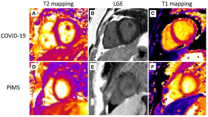
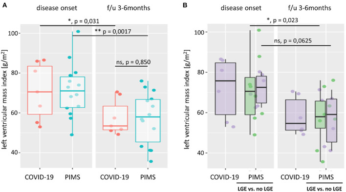
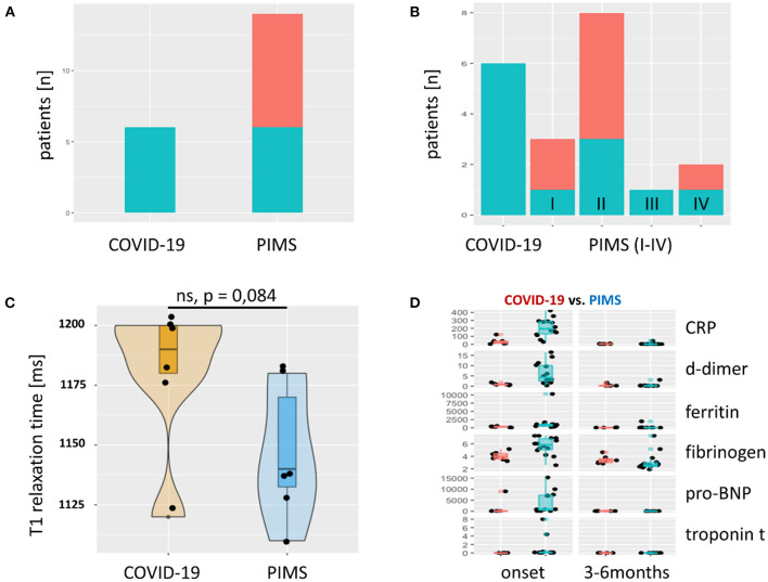
Data were obtained from = 14 patients with PIMS and = 7 patients with severe acute COVID-19. At the start of the respective disease, left ventricular (LV) ejection fraction was reduced in seven patients with PIMS but none in the acute COVID-19 group. Transient mitral valve insufficiency was present in 38% of patients, of whom PIMS accounted for 7/8 cases. Eight patients (38%) with PIMS presented coronary artery abnormalities, with normalization in 7/8 patients. A significant decrease in LV mass index 3-6 months after disease onset was observed in both groups. MRI follow-up revealed non-ischemic myocardial pattern of LGE in 12/21 patients- in all (6/6) after severe acute COVID-19 and in less than half (6/14) after PIMS. Normal body weight-adjusted stroke volumes and end-diastolic volumes were found in 20/21 patients.
We show that children suffering from severe acute COVID-19 have a similar, or worse, cardiac risk profile as patients with PIMS. Both patient groups should therefore receive close pediatric cardiac follow-up examinations. Cardiac MRI is the technique of choice, as most patients presented with delayed LGE as a sign of persistent cardiac injury despite normalization of laboratory and echocardiographic findings.
2019冠状病毒病(COVID-19)在儿童中很少会发展为重症。然而,确实会出现重症病例,尤其是存在风险因素的情况下。少数儿童会发展为具有较高发病率的儿童炎症性多系统综合征(PIMS)。虽然PIMS后心脏受累的重要性已得到充分证实,但其在重症急性COVID-19后的作用仍不清楚。我们旨在使用心脏磁共振成像(MRI)比较重症急性COVID-19后儿童的心脏后遗症,并将其与PIMS后的患者进行比较。
在这项前瞻性队列研究中,我们在单一中心招募了急性COVID或PIMS患者。在疾病发作后2天内以及出院后3至6个月进行临床随访、实验室检查、心电图和超声心动图检查。在3至6个月后的最后一次随访中,进行了带有延迟钆增强(LGE)的心脏MRI(CMR)检查,以评估心脏后遗症并比较两组情况。
共纳入14例PIMS患者和7例重症急性COVID-19患者。在各自疾病开始时,7例PIMS患者的左心室(LV)射血分数降低,而急性COVID-19组中无此情况。38%的患者存在短暂性二尖瓣关闭不全,其中7/8例为PIMS患者。8例(38%)PIMS患者出现冠状动脉异常,7/8例患者恢复正常。两组在疾病发作后3至6个月均观察到左心室质量指数显著下降。MRI随访显示,21例患者中有12例出现非缺血性心肌LGE模式,其中重症急性COVID-19后全部(6/6)出现,PIMS后不到一半(6/14)出现。21例患者中有20例体重调整后的每搏输出量和舒张末期容积正常。
我们表明,患有重症急性COVID-19的儿童与PIMS患者具有相似或更差的心脏风险状况。因此,这两组患者都应接受密切的儿科心脏随访检查。心脏MRI是首选技术,因为尽管实验室检查和超声心动图结果恢复正常,但大多数患者仍出现延迟LGE,这是持续性心脏损伤的迹象。