Guo Guoqiang, Feng Jiaping, Jin Chunchun, Gong Xuehao, Chen Yihao, Chen Sihan, Wei Zhanghong, Xiong Huahua, Lu Jianghao
Department of Ultrasound, Shenzhen Second People's Hospital, The First Affiliated Hospital of Shenzhen University, Sungang West Road 3002, Futian District, Shenzhen 518025, China.
Graduate School, Guangzhou Medical University, Guangzhou 510180, China.
Diagnostics (Basel). 2023 Feb 2;13(3):540. doi: 10.3390/diagnostics13030540.
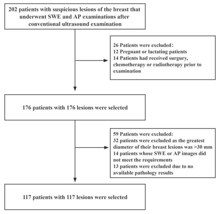
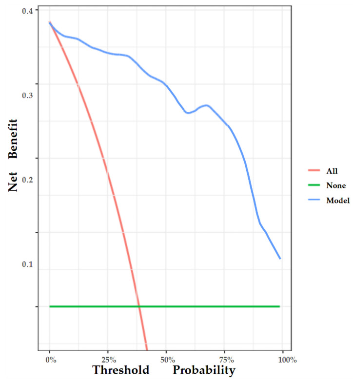
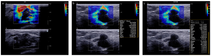
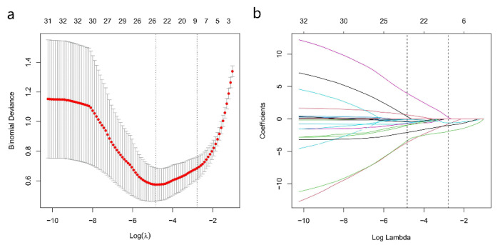
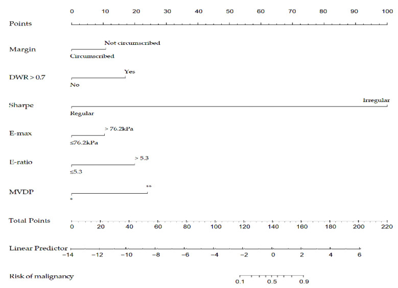
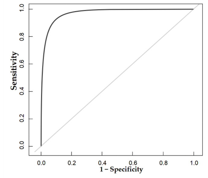
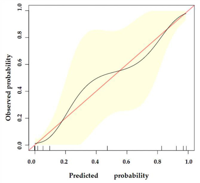
Several studies have demonstrated the difficulties in distinguishing malignant lesions of the breast from benign lesions owing to overlapping morphological features on ultrasound. Consequently, we aimed to develop a nomogram based on shear wave elastography (SWE), Angio Planewave Ultrasensitive imaging (Angio PLUS (AP)), and conventional ultrasound imaging biomarkers to predict malignancy in patients with breast lesions. This prospective study included 117 female patients with suspicious lesions of the breast. Features of lesions were extracted from SWE, AP, and conventional ultrasound images. The least absolute shrinkage and selection operator (Lasso) algorithms were used to select breast cancer-related imaging biomarkers, and a nomogram was developed based on six of the 16 imaging biomarkers. This model exhibited good discrimination (area under the receiver operating characteristic curve (AUC): 0.969; 95% confidence interval (CI): 0.928, 0.989) between malignant and benign breast lesions. Moreover, the nomogram also showed demonstrated good calibration and clinical usefulness. In conclusion, our nomogram can be a potentially useful tool for individually-tailored diagnosis of breast tumors in clinical practice.
多项研究表明,由于超声检查中形态特征重叠,难以区分乳腺恶性病变与良性病变。因此,我们旨在基于剪切波弹性成像(SWE)、血管平面波超声敏感成像(Angio PLUS(AP))和传统超声成像生物标志物开发一种列线图,以预测乳腺病变患者的恶性程度。这项前瞻性研究纳入了117例有乳腺可疑病变的女性患者。从SWE、AP和传统超声图像中提取病变特征。使用最小绝对收缩和选择算子(Lasso)算法选择与乳腺癌相关的成像生物标志物,并基于16个成像生物标志物中的6个开发了列线图。该模型在乳腺恶性病变和良性病变之间表现出良好的区分能力(受试者操作特征曲线下面积(AUC):0.969;95%置信区间(CI):0.928,0.989)。此外,列线图还显示出良好的校准和临床实用性。总之,我们的列线图可能成为临床实践中个体化诊断乳腺肿瘤的有用工具。