Department of Molecular and Comparative Pathobiology, Johns Hopkins University School of Medicine, Baltimore, Maryland.
College of Pharmacy, University of Texas, Houston, Texas, USA.
AIDS. 2023 Apr 1;37(5):733-744. doi: 10.1097/QAD.0000000000003487. Epub 2023 Feb 4.
Latent infection by HIV hinders viral eradication despite effective antiretroviral treatment (ART). Among proposed contributors to viral latency are cellular small RNAs that have also been proposed to shuttle between cells in extracellular vesicles. Thus, we profiled extracellular vesicle small RNAs during different infection phases to understand the potential relationship between these extracellular vesicle associated small RNAs and viral infection.
A well characterized simian immunodeficiency virus (SIV)/macaque model of HIV was used to profile extracellular vesicle enriched blood plasma fractions harvested during preinfection, acute infection, latent infection/ART treatment, and rebound after ART interruption.
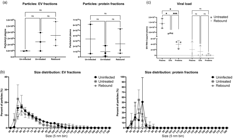
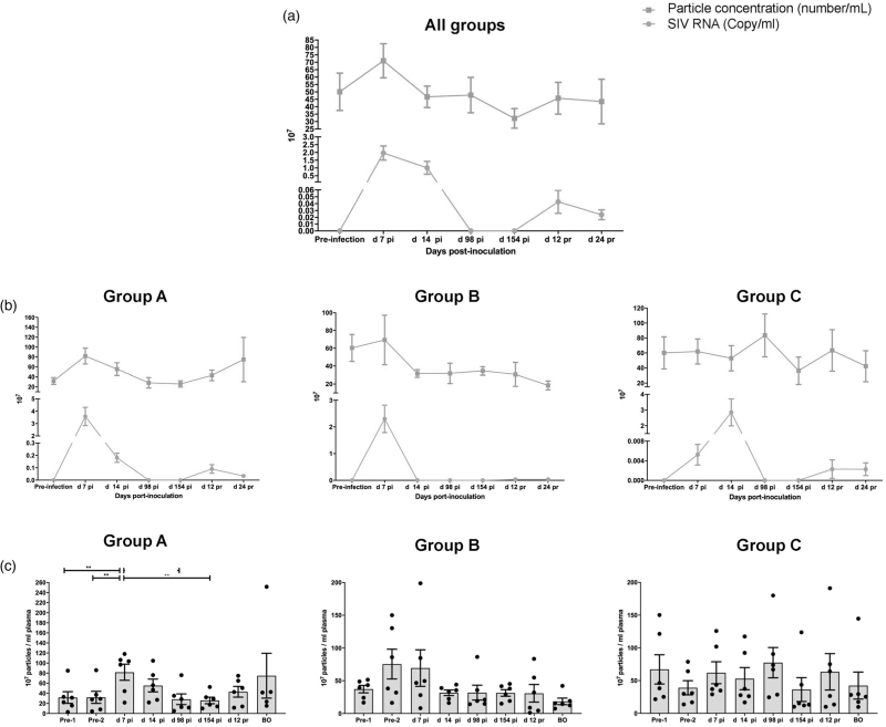
Measurement of extracellular vesicle concentration, size distribution, and morphology was complemented with qPCR array for small RNA expression, followed by individual qPCR validations. Iodixanol density gradients were used to separate extracellular vesicle subtypes and virions.
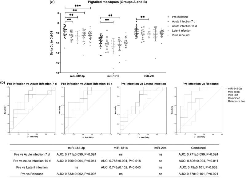
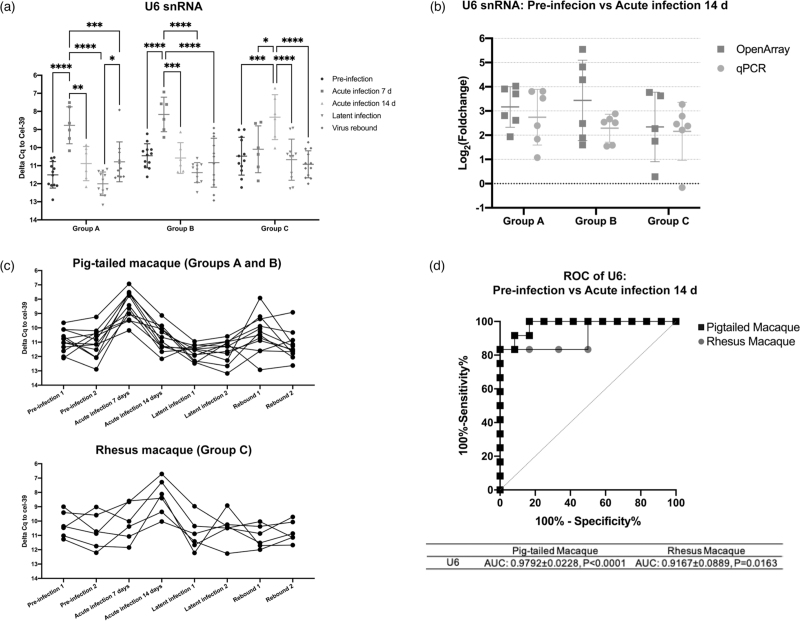
Plasma extracellular vesicle particle counts correlated with viral load and peaked during acute infection. However, SIV gag RNA detection showed that virions did not fully explain this peak. Extracellular vesicle microRNAs miR-181a, miR-342-3p, and miR-29a decreased with SIV infection and remained downregulated in latency. Interestingly, small nuclear RNA U6 had a tight association with viral load peak.
This study is the first to monitor how extracellular vesicle concentration and extracellular vesicle small RNA expression change dynamically in acute viral infection, latency, and rebound in a carefully controlled animal model. These changes may also reveal regulatory roles in retroviral infection and latency.
尽管抗逆转录病毒治疗 (ART) 有效,但 HIV 的潜伏感染仍阻碍了病毒的清除。细胞小 RNA 也被认为在细胞外囊泡中穿梭,是导致病毒潜伏的原因之一。因此,我们在不同的感染阶段对细胞外囊泡小 RNA 进行了分析,以了解这些与细胞外囊泡相关的小 RNA 与病毒感染之间的潜在关系。
使用经过充分验证的猴免疫缺陷病毒 (SIV)/猕猴 HIV 模型,分析在感染前、急性感染、潜伏感染/ART 治疗和 ART 中断后反弹期间采集的富含细胞外囊泡的血浆级分。
用 qPCR 阵列检测小 RNA 的表达,并进行个体 qPCR 验证,以补充细胞外囊泡浓度、大小分布和形态的测量。使用碘克沙醇密度梯度分离细胞外囊泡亚型和病毒粒子。
血浆细胞外囊泡颗粒计数与病毒载量相关,并在急性感染期间达到峰值。然而,SIV gag RNA 的检测表明,病毒粒子并不能完全解释这一峰值。细胞外囊泡 microRNA miR-181a、miR-342-3p 和 miR-29a 的表达随着 SIV 感染而降低,并在潜伏感染中持续下调。有趣的是,小核 RNA U6 与病毒载量峰值密切相关。
本研究首次在精心控制的动物模型中监测急性病毒感染、潜伏和反弹期间细胞外囊泡浓度和细胞外囊泡小 RNA 表达的动态变化。这些变化也可能揭示了逆转录病毒感染和潜伏的调控作用。