Li Lucie Y, Kreye Jakob, Burek Malgorzata, Cordero-Gomez César, Barthel Paula C, Sánchez-Sendín Elisa, Kornau Hans-Christian, Schmitz Dietmar, Scharf Madeleine, Meybohm Patrick, Reincke S Momsen, Prüss Harald, Höltje Markus
Institute of Integrative Neuroanatomy Berlin, Charité-Universitätsmedizin Berlin, corporate member of Freie Universität Berlin, Humboldt-Universität zu Berlin and Berlin Institute of Health, Berlin, Germany.
German Center for Neurodegenerative Diseases (DZNE) Berlin, Berlin, Germany.
Front Cell Neurosci. 2023 Jan 30;17:1077204. doi: 10.3389/fncel.2023.1077204. eCollection 2023.
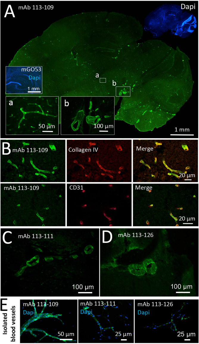
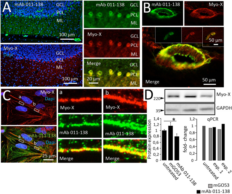
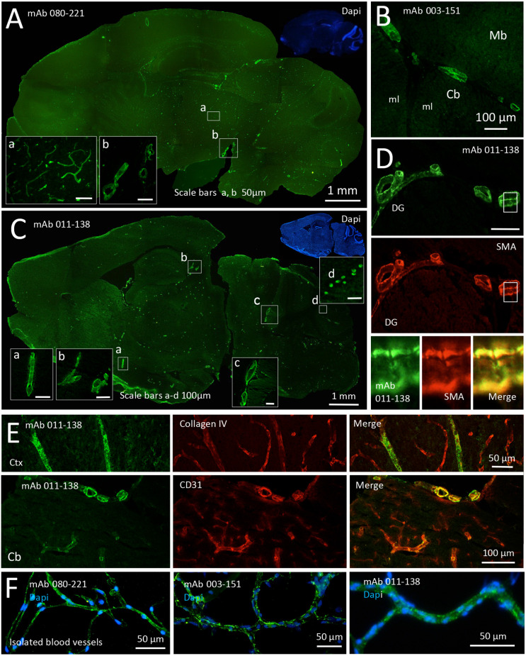
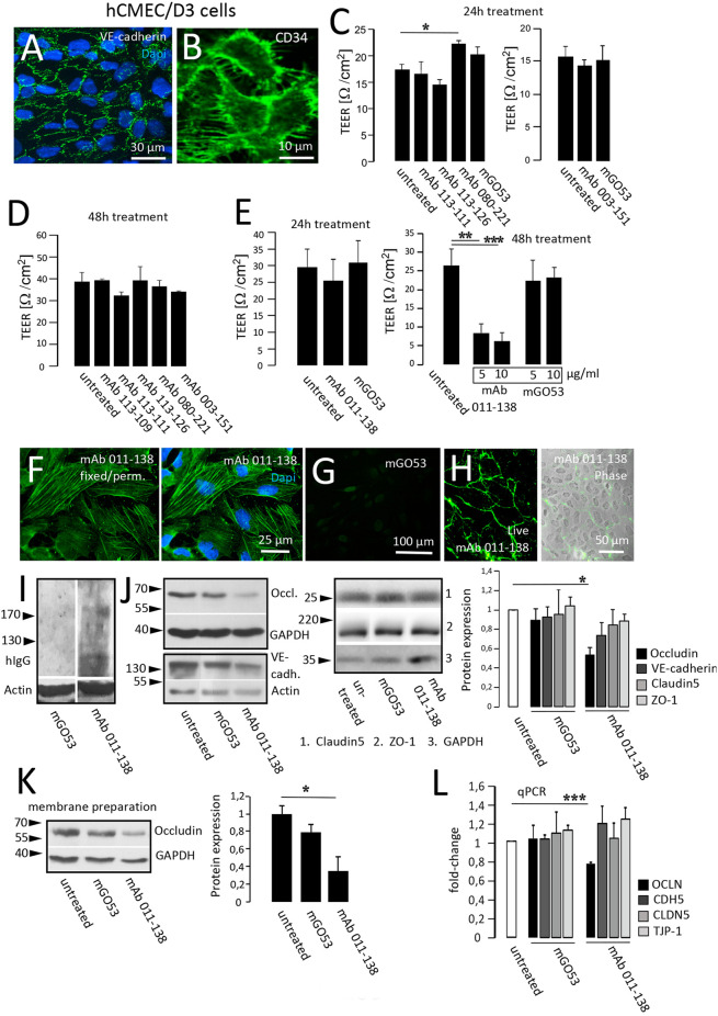
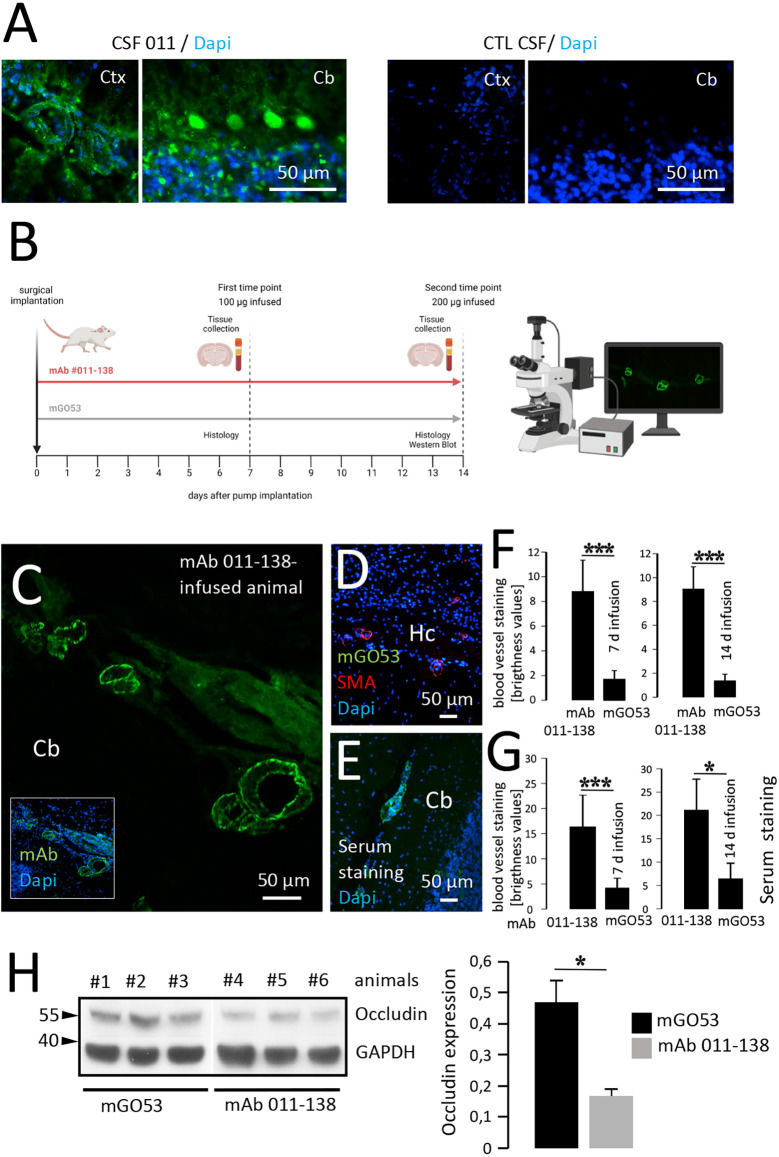
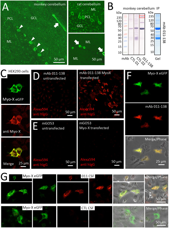
The antibody repertoire from CSF-derived antibody-secreting cells and memory B-cells in patients with encephalitis contains a considerable number of antibodies that do not target the disease-defining autoantigen such as the GABA or NMDA receptors. This study focuses on the functional relevance of autoantibodies to brain blood vessels in patients with GABA and NMDA receptor encephalitis. We tested 149 human monoclonal IgG antibodies from the cerebrospinal fluid of six patients with different forms of autoimmune encephalitis on murine brain sections for reactivity to blood vessels using immunohistochemistry. Positive candidates were tested for reactivity with purified brain blood vessels, effects on transendothelial electrical resistance (TEER), and expression of tight junction proteins as well as gene regulation using human brain microvascular endothelial hCMEC/D3 cells as blood-brain barrier model. One blood-vessel reactive antibody was infused intrathecally by pump injection in mice to study binding and effects on tight junction proteins such as Occludin. Target protein identification was addressed using transfected HEK293 cells. Six antibodies reacted with brain blood vessels, three were from the same patient with GABAR encephalitis, and the other three were from different patients with NMDAR encephalitis. One antibody from an NMDAR encephalitis patient, mAb 011-138, also reacted with cerebellar Purkinje cells. In this case, treatment of hCMEC/D3 cells resulted in decreased TEER, reduced Occludin expression, and mRNA levels. Functional relevance was confirmed as Occludin downregulation was observed in mAb 011-138-infused animals. Unconventional Myosin-X was identified as a novel autoimmune target for this antibody. We conclude that autoantibodies to blood vessels occur in autoimmune encephalitis patients and might contribute to a disruption of the blood-brain barrier thereby suggesting a potential pathophysiological relevance of these antibodies.
脑炎患者脑脊液来源的抗体分泌细胞和记忆B细胞中的抗体库包含大量不靶向疾病定义自身抗原(如GABA或NMDA受体)的抗体。本研究聚焦于GABA和NMDA受体脑炎患者中自身抗体与脑血 管的功能相关性。我们使用免疫组化方法,在小鼠脑切片上检测了来自6例不同形式自身免疫性脑炎患者脑脊液中的149种人单克隆IgG抗体与血管的反应性。对阳性候选抗体进行了与纯化脑血 管的反应性测试、对跨内皮电阻(TEER)的影响、紧密连接蛋白的表达以及使用人脑微血管内皮hCMEC/D3细胞作为血脑屏障模型的基因调控测试。通过泵注射将一种与血管反应的抗体鞘内注射到小鼠体内,以研究其与紧密连接蛋白(如闭合蛋白)的结合及影响。使用转染的HEK293细胞进行靶蛋白鉴定。六种抗体与脑血 管反应,三种来自同一例GABAR脑炎患者,另外三种来自不同的NMDAR脑炎患者。来自一名NMDAR脑炎患者的一种抗体mAb 011 - 138也与小脑浦肯野细胞反应。在这种情况下,对hCMEC/D3细胞的处理导致TEER降低、闭合蛋白表达减少以及mRNA水平降低。在注射mAb 011 - 138的动物中观察到闭合蛋白下调,证实了其功能相关性。非传统肌球蛋白X被鉴定为该抗体的一种新型自身免疫靶点。我们得出结论,自身免疫性脑炎患者中存在针对血管的自身抗体,可能导致血脑屏障破坏,从而提示这些抗体具有潜在的病理生理相关性。