• 文献检索
  • 文档翻译
  • 深度研究
  • 学术资讯
  • Suppr Zotero 插件Zotero 插件
  • 邀请有礼
  • 套餐&价格
  • 历史记录
应用&插件
Suppr Zotero 插件Zotero 插件浏览器插件Mac 客户端Windows 客户端微信小程序
定价
高级版会员购买积分包购买API积分包
服务
文献检索文档翻译深度研究API 文档MCP 服务
关于我们
关于 Suppr公司介绍联系我们用户协议隐私条款
关注我们

Suppr 超能文献

核心技术专利:CN118964589B侵权必究
粤ICP备2023148730 号-1Suppr @ 2026

文献检索

告别复杂PubMed语法,用中文像聊天一样搜索,搜遍4000万医学文献。AI智能推荐,让科研检索更轻松。

立即免费搜索

文件翻译

保留排版,准确专业,支持PDF/Word/PPT等文件格式,支持 12+语言互译。

免费翻译文档

深度研究

AI帮你快速写综述,25分钟生成高质量综述,智能提取关键信息,辅助科研写作。

立即免费体验

心肌细胞与成纤维细胞的相互作用调节心肌损伤后的铁死亡和纤维化。

Cardiomyocyte-fibroblast interaction regulates ferroptosis and fibrosis after myocardial injury.

作者信息

Mohr Mary E, Li Shuang, Trouten Allison M, Stairley Rebecca A, Roddy Patrick L, Liu Chun, Zhang Min, Sucov Henry M, Tao Ge

机构信息

Department of Regenerative Medicine and Cell Biology, Medical University of South Carolina, Charleston, SC 29425, USA.

These authors contributed equally.

出版信息

bioRxiv. 2023 Feb 8:2023.02.07.527364. doi: 10.1101/2023.02.07.527364.

DOI:10.1101/2023.02.07.527364
PMID:36798323
原文链接:https://pmc.ncbi.nlm.nih.gov/articles/PMC9934560/
Abstract

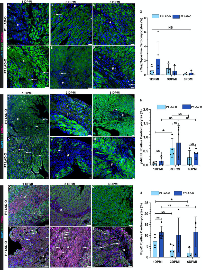
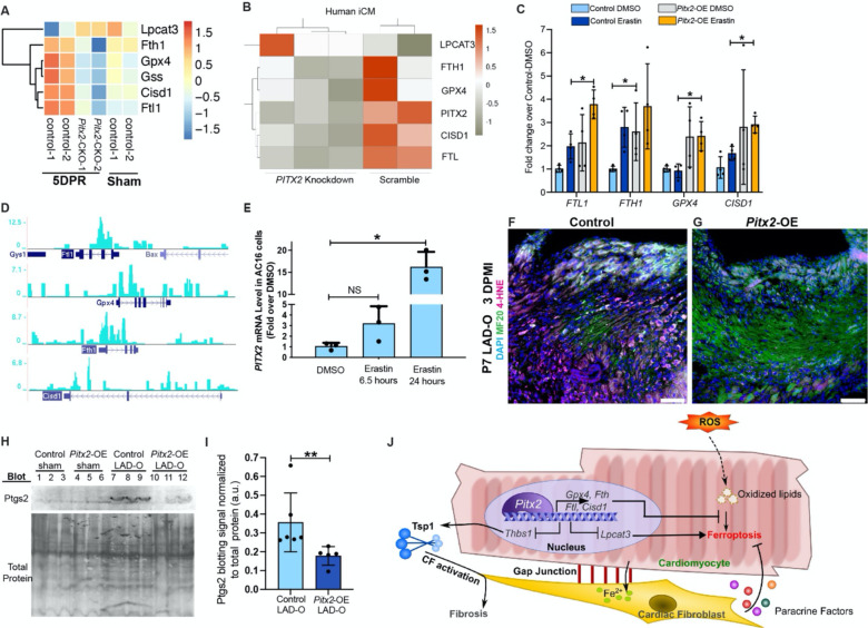
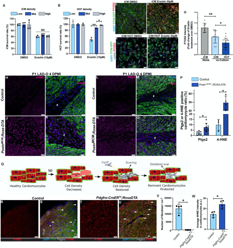
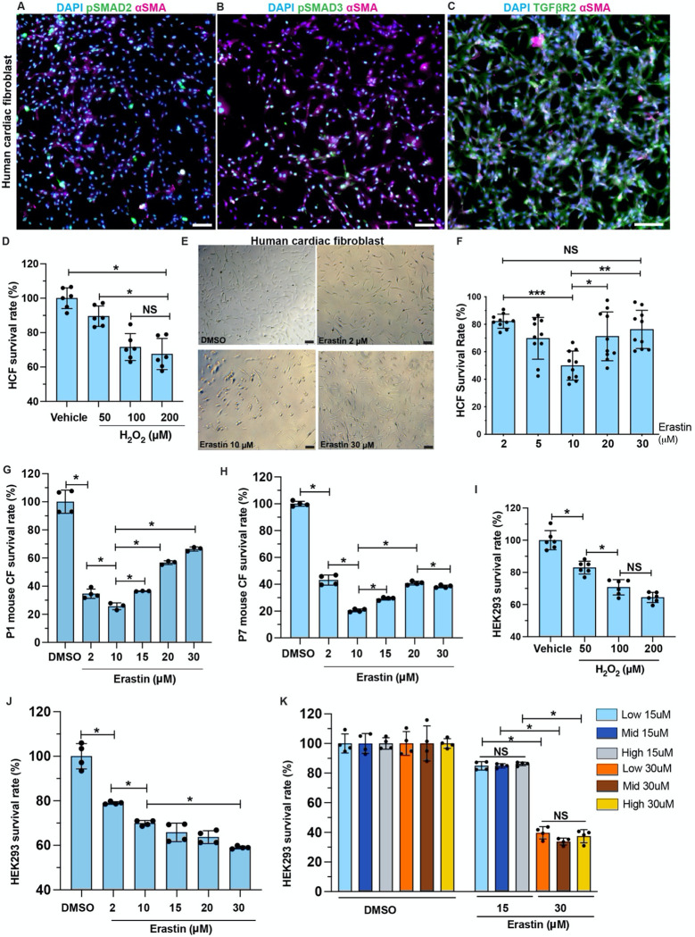
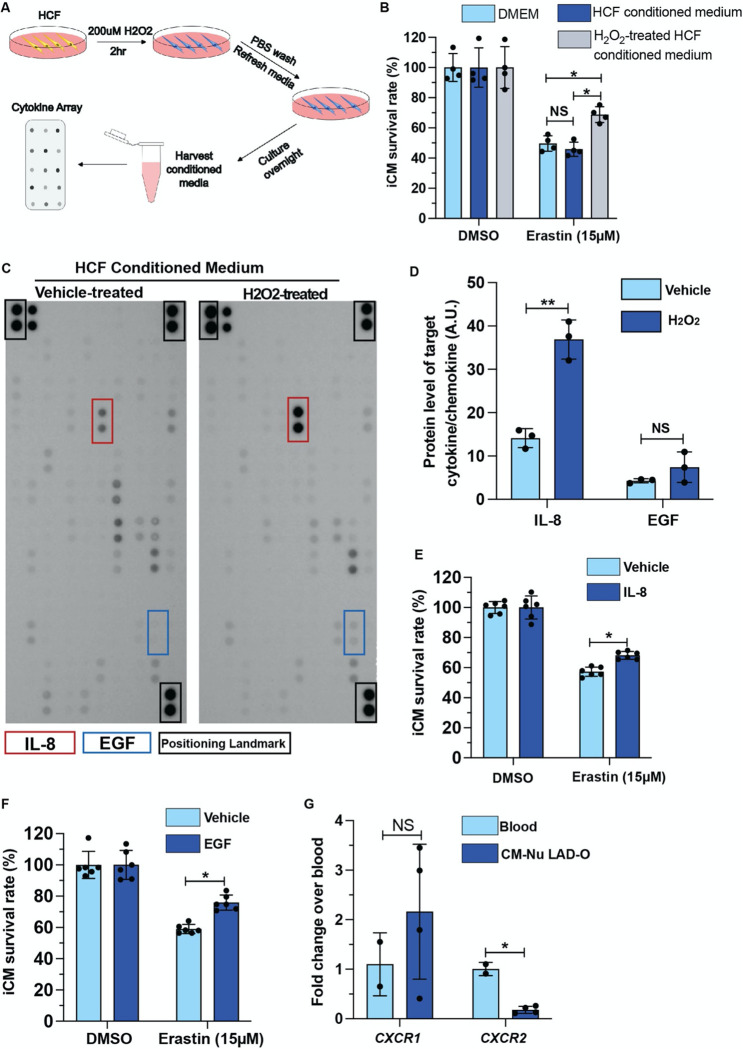
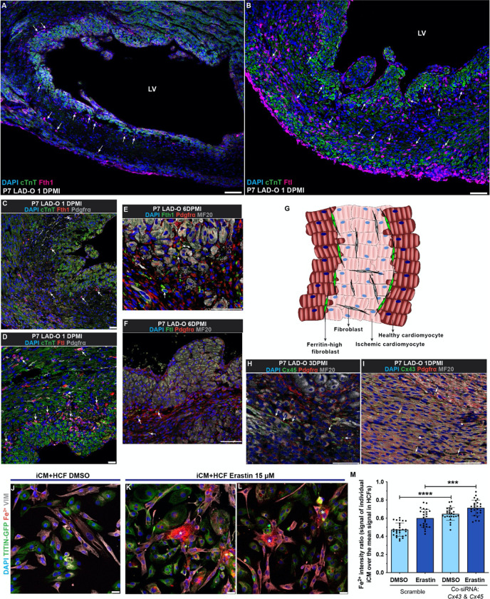
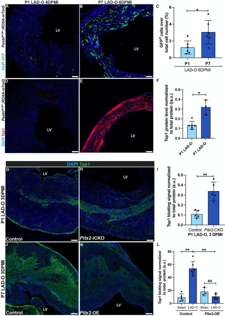
Neonatal mouse hearts have transient renewal capacity which is lost in juvenile and adult hearts. After myocardial infarction (MI) in neonatal hearts, an initial loss of cardiomyocytes occurs but it is unclear through which type of regulated cell death (RCD). In the current studies, we induced MI in neonatal and juvenile mouse hearts, and show that ischemic cardiomyocytes primarily undergo ferroptosis, a non-apoptotic and iron-dependent form of RCD. We demonstrate that cardiac fibroblasts (CFs) protect cardiomyocytes from ferroptosis through paracrine factors and direct cell-cell interaction. CFs show strong resistance to ferroptosis due to high ferritin expression. Meanwhile, the fibrogenic role of CFs, typically considered detrimental to heart function, is negatively regulated by paired-like homeodomain 2 (Pitx2) signaling from cardiomyocytes. In addition, Pitx2 prevents ferroptosis in cardiomyocytes by regulating ferroptotic genes. Understanding the regulatory mechanisms of cardiomyocyte survival and death can identify potentially translatable therapeutic strategies for MI.

摘要

新生小鼠心脏具有短暂的更新能力,而这种能力在幼年和成年心脏中会丧失。在新生心脏发生心肌梗死(MI)后,会出现心肌细胞的初始丧失,但尚不清楚是通过哪种类型的程序性细胞死亡(RCD)。在当前的研究中,我们在新生和幼年小鼠心脏中诱导MI,并表明缺血心肌细胞主要经历铁死亡,这是一种非凋亡且依赖铁的RCD形式。我们证明心脏成纤维细胞(CFs)通过旁分泌因子和直接的细胞间相互作用保护心肌细胞免受铁死亡。由于铁蛋白高表达,CFs对铁死亡表现出强大的抗性。同时,通常被认为对心脏功能有害的CFs的纤维化作用受到心肌细胞中配对样同源结构域2(Pitx2)信号通路的负调控。此外,Pitx2通过调节铁死亡基因来防止心肌细胞发生铁死亡。了解心肌细胞存活和死亡的调控机制可以确定针对MI的潜在可转化治疗策略。

https://cdn.ncbi.nlm.nih.gov/pmc/blobs/784a/9934560/bf205e1ac484/nihpp-2023.02.07.527364v1-f0007.jpg
https://cdn.ncbi.nlm.nih.gov/pmc/blobs/784a/9934560/db7f64bb3dcc/nihpp-2023.02.07.527364v1-f0001.jpg
https://cdn.ncbi.nlm.nih.gov/pmc/blobs/784a/9934560/cc6252ab5d3e/nihpp-2023.02.07.527364v1-f0002.jpg
https://cdn.ncbi.nlm.nih.gov/pmc/blobs/784a/9934560/41e9922cd458/nihpp-2023.02.07.527364v1-f0003.jpg
https://cdn.ncbi.nlm.nih.gov/pmc/blobs/784a/9934560/4c3e8724e3d0/nihpp-2023.02.07.527364v1-f0004.jpg
https://cdn.ncbi.nlm.nih.gov/pmc/blobs/784a/9934560/9853001e3fbb/nihpp-2023.02.07.527364v1-f0005.jpg
https://cdn.ncbi.nlm.nih.gov/pmc/blobs/784a/9934560/3f1e7a5a19ad/nihpp-2023.02.07.527364v1-f0006.jpg
https://cdn.ncbi.nlm.nih.gov/pmc/blobs/784a/9934560/bf205e1ac484/nihpp-2023.02.07.527364v1-f0007.jpg

相似文献

1
Cardiomyocyte-fibroblast interaction regulates ferroptosis and fibrosis after myocardial injury.心肌细胞与成纤维细胞的相互作用调节心肌损伤后的铁死亡和纤维化。
bioRxiv. 2023 Feb 8:2023.02.07.527364. doi: 10.1101/2023.02.07.527364.
2
Cardiomyocyte-fibroblast interaction regulates ferroptosis and fibrosis after myocardial injury.心肌细胞与成纤维细胞的相互作用调节心肌损伤后的铁死亡和纤维化。
iScience. 2024 Feb 15;27(3):109219. doi: 10.1016/j.isci.2024.109219. eCollection 2024 Mar 15.
3
Anti-Ferroptotic Treatment Deteriorates Myocardial Infarction by Inhibiting Angiogenesis and Altering Immune Response.抗铁死亡治疗通过抑制血管生成和改变免疫反应使心肌梗死恶化。
Antioxidants (Basel). 2024 Jun 26;13(7):769. doi: 10.3390/antiox13070769.
4
Salvia miltiorrhiza suppresses cardiomyocyte ferroptosis after myocardial infarction by activating Nrf2 signaling.丹参通过激活 Nrf2 信号通路抑制心肌梗死后心肌细胞铁死亡。
J Ethnopharmacol. 2024 Aug 10;330:118214. doi: 10.1016/j.jep.2024.118214. Epub 2024 Apr 17.
5
Exosomes secreted from cardiomyocytes suppress the sensitivity of tumor ferroptosis in ischemic heart failure.心肌细胞分泌的外泌体抑制缺血性心力衰竭中肿瘤细胞的铁死亡敏感性。
Signal Transduct Target Ther. 2023 Mar 27;8(1):121. doi: 10.1038/s41392-023-01336-4.
6
Single-cell sequencing reveals LRRc17-mediated modulation of cardiac fibroblast ferroptosis in regulating myocardial fibrosis after myocardial infarction.单细胞测序揭示 LRRc17 介导的心脏成纤维细胞铁死亡调控在心肌梗死后心肌纤维化中的作用。
Cell Signal. 2024 Sep;121:111293. doi: 10.1016/j.cellsig.2024.111293. Epub 2024 Jul 10.
7
Adaptor protein HIP-55-mediated signalosome protects against ferroptosis in myocardial infarction.衔接蛋白 HIP-55 介导的信号复合物保护心肌梗死中的铁死亡。
Cell Death Differ. 2023 Mar;30(3):825-838. doi: 10.1038/s41418-022-01110-z. Epub 2023 Jan 13.
8
Nrf3-Mediated Mitochondrial Superoxide Promotes Cardiomyocyte Apoptosis and Impairs Cardiac Functions by Suppressing Pitx2.Nrf3介导的线粒体超氧化物通过抑制Pitx2促进心肌细胞凋亡并损害心脏功能。
Circulation. 2025 Apr 8;151(14):1024-1046. doi: 10.1161/CIRCULATIONAHA.124.070286. Epub 2025 Mar 18.
9
Ferroptotic cardiomyocyte-derived exosomes promote cardiac macrophage M1 polarization during myocardial infarction.铁死亡性心肌细胞来源的外泌体在心肌梗死后促进心肌巨噬细胞 M1 极化。
PeerJ. 2022 Jul 6;10:e13717. doi: 10.7717/peerj.13717. eCollection 2022.
10
Cardiac Fibroblasts and Myocardial Regeneration.心脏成纤维细胞与心肌再生
Front Bioeng Biotechnol. 2021 Mar 25;9:599928. doi: 10.3389/fbioe.2021.599928. eCollection 2021.