Department of Cardiology, Huashan Hospital, Fudan University, Shanghai, China.
Nursing Department, Huashan Hospital, Fudan University, Shanghai, China.
PeerJ. 2022 Jul 6;10:e13717. doi: 10.7717/peerj.13717. eCollection 2022.
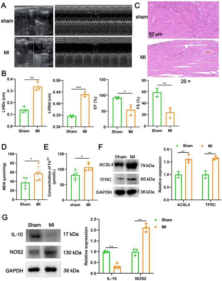
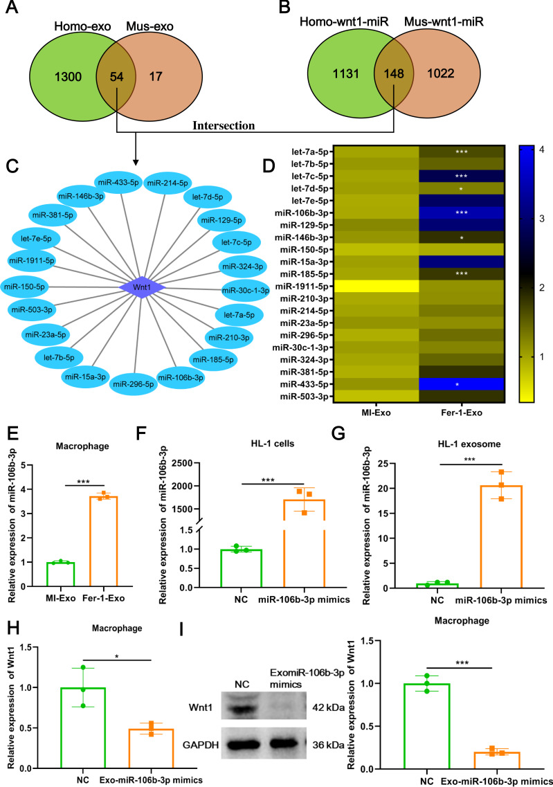
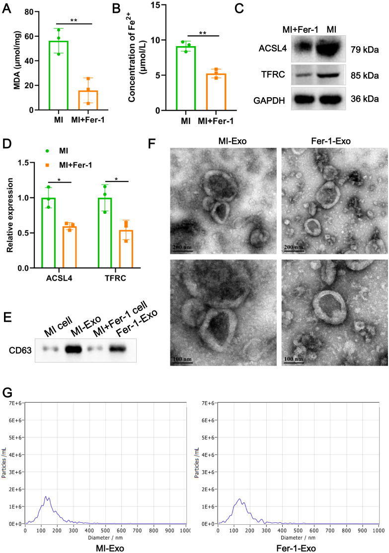
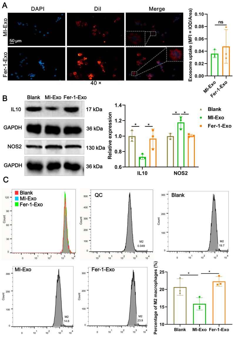
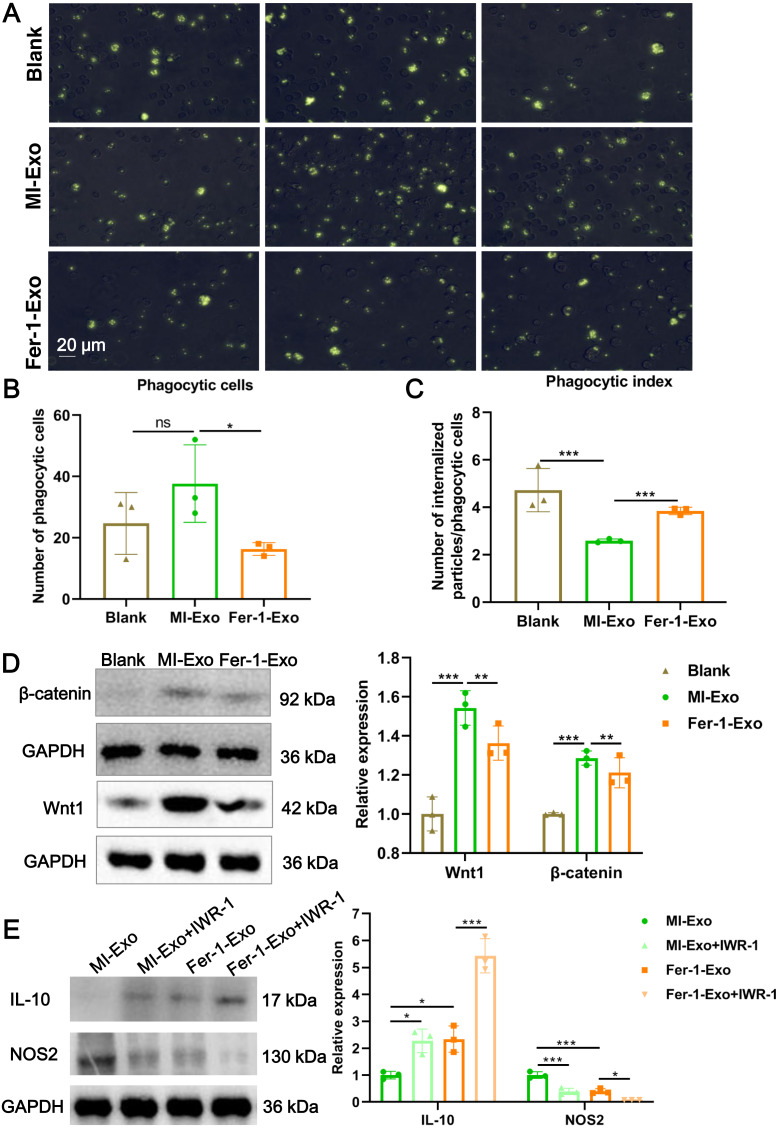
Ferroptosis is a mode of cell death that occurs in myocardial infarction (MI). Signals emanating from apoptotic cells are able to induce macrophage polarization through exosome-loading cargos, which plays a vital role in the process of disease. However, whether ferroptotic cardiomyocytes derived exosome (MI-Exo) during MI act on macrophage polarization and its mechanism remain unclear. In this study, a MI mouse model was established, and cardiac function evaluation and pathological staining were performed. The effect of MI-Exo on polarization of RAW264.7 cells was assessed by the expression of IL-10 and NOS2. Ferroptosis inhibitor of ferrostatin-1 was used to verify whether MI-Exo function was dependents on ferroptosis. Cardiac function and myocardial histomorphology were markedly impaired and massive immune cell infiltration in MI mice, compared with the sham group. The significantly increased MDA content and Fe accumulation in the heart tissue of MI mice suggested cardiomyocyte ferroptosis. Compared with the sham group, the expression of M1 marker NOS2 was significantly up-regulated and M2 marker IL-10 was significantly down-regulated in the heart tissue of MI mice. Exosome-derived from MI HL-1 cell-treated with ferrostatin-1 (Fer-1-Exo) and MI-Exo were internalized by RAW 264.7 cells. Compared with culture alone, co-cultured with MI-Exo significantly promoted NOS2 expression and suppressed IL-10 expression, and decreased proportion of Arginase-1-labeled M2 macrophages, also inhibited phagocytosis of RAW 264.7 cells. Wnt1 and β-cantenin expression also elevated after treated with MI-Exo. However, co-cultured with Fer-1-Exo significantly reversed the above changes on RAW 264.7 cells induced by MI-Exo. In conclusion, ferroptotic cardiomyocytes-derived exosome crosstalk macrophage to induce M1 polarization via Wnt/β-cantenin pathway, resulting in pathological progress in MI. This understanding provides novel therapeutic target for MI.
铁死亡是心肌梗死 (MI) 中发生的一种细胞死亡方式。凋亡细胞发出的信号能够通过外体装载货物诱导巨噬细胞极化,这在疾病过程中起着至关重要的作用。然而,MI 期间铁死亡心肌细胞衍生的外体 (MI-Exo) 是否作用于巨噬细胞极化及其机制尚不清楚。在这项研究中,建立了 MI 小鼠模型,并进行了心脏功能评估和病理染色。通过 IL-10 和 NOS2 的表达来评估 MI-Exo 对 RAW264.7 细胞极化的影响。使用铁死亡抑制剂 ferrostatin-1 来验证 MI-Exo 的功能是否依赖于铁死亡。与假手术组相比,MI 小鼠的心脏功能和心肌组织形态学明显受损,大量免疫细胞浸润。MI 小鼠心脏组织中 MDA 含量和 Fe 积累的显著增加表明心肌细胞发生铁死亡。与假手术组相比,MI 小鼠心脏组织中 M1 标志物 NOS2 的表达显著上调,M2 标志物 IL-10 的表达显著下调。用 ferrostatin-1(Fer-1-Exo)处理的 MI HL-1 细胞衍生的外体和 MI-Exo 被 RAW 264.7 细胞内化。与单独培养相比,与 MI-Exo 共培养显著促进了 NOS2 的表达,抑制了 IL-10 的表达,减少了 Arg1 标记的 M2 巨噬细胞的比例,也抑制了 RAW 264.7 细胞的吞噬作用。用 MI-Exo 处理后,Wnt1 和 β-cantenin 的表达也升高。然而,与 Fer-1-Exo 共培养显著逆转了 MI-Exo 对 RAW 264.7 细胞的上述变化。总之,铁死亡心肌细胞衍生的外体通过 Wnt/β-cantenin 途径与巨噬细胞相互作用,诱导 M1 极化,导致 MI 的病理进展。这一认识为 MI 提供了新的治疗靶点。