• 文献检索
  • 文档翻译
  • 深度研究
  • 学术资讯
  • Suppr Zotero 插件Zotero 插件
  • 邀请有礼
  • 套餐&价格
  • 历史记录
应用&插件
Suppr Zotero 插件Zotero 插件浏览器插件Mac 客户端Windows 客户端微信小程序
定价
高级版会员购买积分包购买API积分包
服务
文献检索文档翻译深度研究API 文档MCP 服务
关于我们
关于 Suppr公司介绍联系我们用户协议隐私条款
关注我们

Suppr 超能文献

核心技术专利:CN118964589B侵权必究
粤ICP备2023148730 号-1Suppr @ 2026

文献检索

告别复杂PubMed语法,用中文像聊天一样搜索,搜遍4000万医学文献。AI智能推荐,让科研检索更轻松。

立即免费搜索

文件翻译

保留排版,准确专业,支持PDF/Word/PPT等文件格式,支持 12+语言互译。

免费翻译文档

深度研究

AI帮你快速写综述,25分钟生成高质量综述,智能提取关键信息,辅助科研写作。

立即免费体验

未治疗的胸腺癌患者的预后 CT 特征。

Prognostic CT features in patients with untreated thymic epithelial tumors.

机构信息

Department of Medical Imaging, Huizhou Municipal Central Hospital, No. 41, North Eling Road, Huizhou, 516001, People's Republic of China.

Department of Radiology, Shandong Tumor Hospital, No.44, Jiyan Road, Jinan, 250117, People's Republic of China.

出版信息

Sci Rep. 2023 Feb 19;13(1):2910. doi: 10.1038/s41598-023-30041-z.

DOI:10.1038/s41598-023-30041-z
PMID:36801902
原文链接:https://pmc.ncbi.nlm.nih.gov/articles/PMC9939415/
Abstract

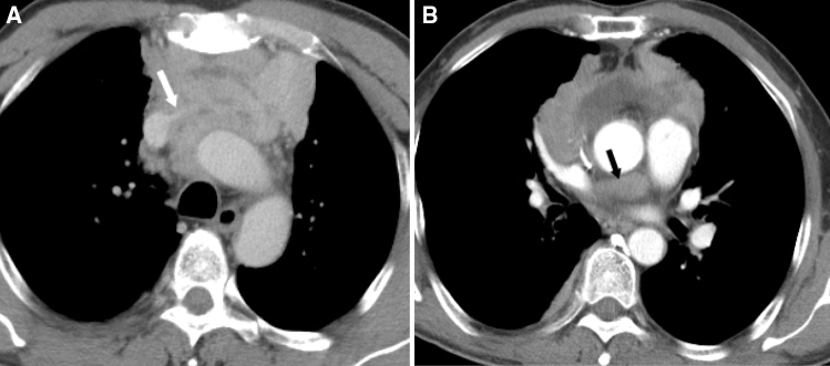
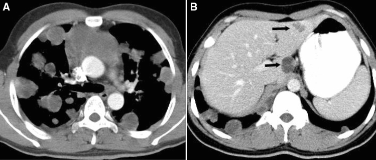
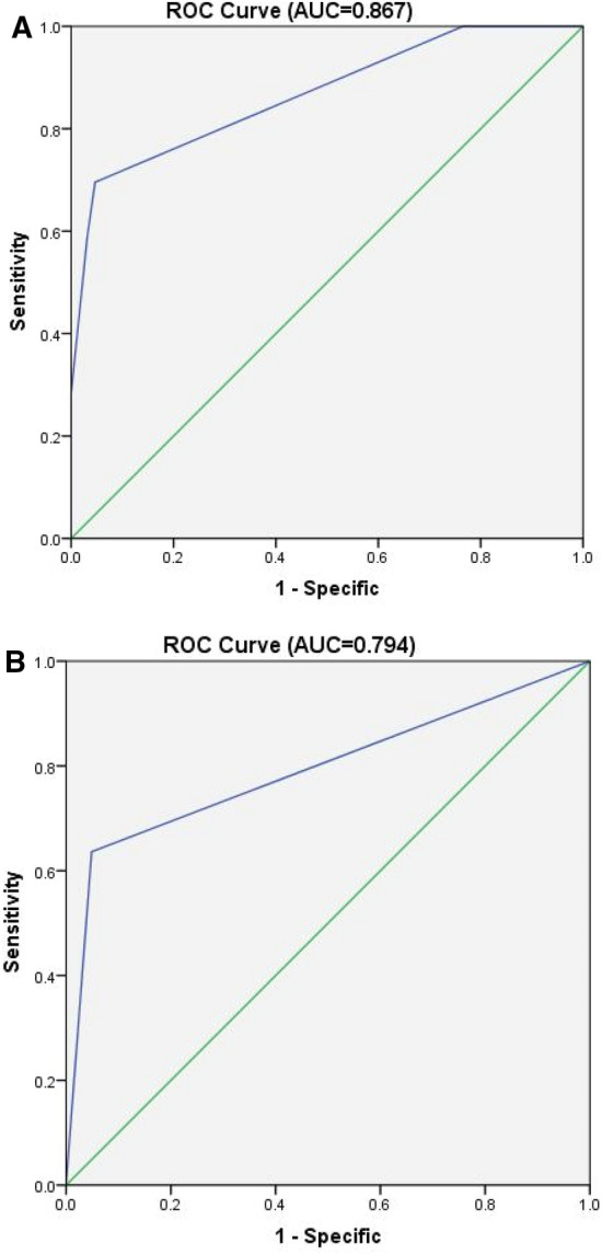
To determine the prognostic CT features in patients with untreated thymic epithelial tumors (TETs). Clinical data and CT imaging features of 194 patients with pathologically confirmed TETs were retrospectively reviewed. The subjects included 113 male and 81 female patients between 15 and 78 years of age, with a mean age of 53.8 years. Clinical outcomes were categorized according to whether relapse, metastasis or death occurred within 3 years after the first diagnosis. Associations between clinical outcomes and CT imaging features were determined using univariate and multivariate logistic regression analyses, while the survival status was analyzed by Cox regression. In this study, we analyzed 110 thymic carcinomas, 52 high-risk thymomas and 32 low-risk thymomas. Percentages of poor outcome and patient death in thymic carcinomas were much higher than those in patients with high-risk and low-risk thymomas. In the thymic carcinomas groups, 46 patients (41.8%) experienced tumor progression, local relapse or metastasis and were categorized as having poor outcomes; vessel invasion and pericardial mass were confirmed to be independent predictors by logistic regression analysis (p < 0.01). In the high-risk thymoma group, 11 patients (21.2%) were categorized as having poor outcomes, and the CT feature pericardial mass was confirmed to be an independent predictor (p < 0.01). In survival analysis, Cox regression showed that CT features of lung invasion, great vessel invasion, lung metastasis and distant organ metastasis were independent predictors for worse survival in the thymic carcinoma group (p < 0.01), while lung invasion and pericardial mass were independent predictors for worse survival in high-risk thymoma group. No CT features were related to poor outcome and worse survival in the low-risk thymoma group. Patients with thymic carcinoma had poorer prognosis and worse survival than those with high-risk or low-risk thymoma. CT can serve as an important tool for predicting the prognosis and survival of patients with TETs. In this cohort, CT features of vessel invasion and pericardial mass were related to poorer outcomes in those with thymic carcinoma and pericardial mass in those with high-risk thymoma. Features including lung invasion, great vessel invasion, lung metastasis and distant organ metastasis indicate worse survival in thymic carcinoma, whereas lung invasion and pericardial mass indicate worse survival in high-risk thymoma.

摘要

为了确定未经治疗的胸腺瘤患者的预后 CT 特征。回顾性分析了 194 例经病理证实的胸腺瘤患者的临床资料和 CT 影像学特征。受试者包括 113 名男性和 81 名女性,年龄在 15 至 78 岁之间,平均年龄为 53.8 岁。根据首次诊断后 3 年内是否复发、转移或死亡,将临床结局分为复发、转移或死亡组和无复发、转移或死亡组。采用单因素和多因素逻辑回归分析比较两组 CT 影像学特征的差异,采用 COX 回归分析生存状态。本研究共分析了 110 例胸腺癌、52 例高危胸腺瘤和 32 例低危胸腺瘤。胸腺癌患者的不良预后和死亡比例明显高于高危和低危胸腺瘤患者。在胸腺癌组中,46 例(41.8%)患者发生肿瘤进展、局部复发或转移,预后不良;血管侵犯和心包肿块被证实是独立的预测因素(p<0.01)。在高危胸腺瘤组中,11 例(21.2%)患者预后不良,心包肿块是独立的预测因素(p<0.01)。在生存分析中,COX 回归显示 CT 特征肺侵犯、大血管侵犯、肺转移和远处器官转移是胸腺癌患者生存不良的独立预测因素(p<0.01),而肺侵犯和心包肿块是高危胸腺瘤患者生存不良的独立预测因素。低危胸腺瘤组 CT 特征与不良预后和生存不良无关。胸腺癌患者的预后和生存明显差于高危和低危胸腺瘤患者。CT 可作为预测胸腺瘤患者预后和生存的重要工具。在本队列中,血管侵犯和心包肿块与胸腺癌较差的预后有关,而心包肿块与高危胸腺瘤的预后有关。肺侵犯、大血管侵犯、肺转移和远处器官转移等 CT 特征提示胸腺癌患者的生存较差,而肺侵犯和心包肿块则提示高危胸腺瘤患者的生存较差。

https://cdn.ncbi.nlm.nih.gov/pmc/blobs/d6e6/9939415/635b0be9f64f/41598_2023_30041_Fig4_HTML.jpg
https://cdn.ncbi.nlm.nih.gov/pmc/blobs/d6e6/9939415/f5af8f0ca01f/41598_2023_30041_Fig1_HTML.jpg
https://cdn.ncbi.nlm.nih.gov/pmc/blobs/d6e6/9939415/6ad4fef2b489/41598_2023_30041_Fig2_HTML.jpg
https://cdn.ncbi.nlm.nih.gov/pmc/blobs/d6e6/9939415/ba84786a33c0/41598_2023_30041_Fig3_HTML.jpg
https://cdn.ncbi.nlm.nih.gov/pmc/blobs/d6e6/9939415/635b0be9f64f/41598_2023_30041_Fig4_HTML.jpg

相似文献

1
Prognostic CT features in patients with untreated thymic epithelial tumors.未治疗的胸腺癌患者的预后 CT 特征。
Sci Rep. 2023 Feb 19;13(1):2910. doi: 10.1038/s41598-023-30041-z.
2
Does CT of thymic epithelial tumors enable us to differentiate histologic subtypes and predict prognosis?胸腺上皮肿瘤的CT检查能否让我们区分组织学亚型并预测预后?
AJR Am J Roentgenol. 2004 Aug;183(2):283-9. doi: 10.2214/ajr.183.2.1830283.
3
Thymic epithelial tumors: comparison of CT and MR imaging findings of low-risk thymomas, high-risk thymomas, and thymic carcinomas.胸腺上皮肿瘤:低危胸腺瘤、高危胸腺瘤和胸腺癌的CT与MR成像表现比较
Eur J Radiol. 2006 Oct;60(1):70-9. doi: 10.1016/j.ejrad.2006.05.003.
4
Clinical characteristics, prognostic factors, and long-term outcomes associated with epithelial malignancies of the thymus: A 20-year single-institution experience.胸腺上皮性恶性肿瘤的临床特征、预后因素及长期预后:20 年单中心经验。
Cancer Rep (Hoboken). 2023 Mar;6(3):e1750. doi: 10.1002/cnr2.1750. Epub 2022 Nov 12.
5
Correlation of clinical and computed tomography features of thymic epithelial tumours with World Health Organization classification and Masaoka-Koga staging.胸内上皮性肿瘤的临床和 CT 特征与世界卫生组织分类和 Masaoka-Koga 分期的相关性。
Eur J Cardiothorac Surg. 2022 Mar 24;61(4):742-748. doi: 10.1093/ejcts/ezab349.
6
Computed tomography radiomic feature analysis of thymic epithelial tumors: Differentiation of thymic epithelial tumors from thymic cysts and prediction of histological subtypes.计算机断层扫描放射组学特征分析胸腺瘤:胸腺瘤与胸囊肿的鉴别及组织学分型预测。
Jpn J Radiol. 2024 Apr;42(4):367-373. doi: 10.1007/s11604-023-01512-0. Epub 2023 Nov 27.
7
Predictive Features of Thymic Carcinoma and High-Risk Thymomas Using Random Forest Analysis.使用随机森林分析的胸腺癌和高危胸腺瘤的预测特征
J Comput Assist Tomogr. 2020 Nov/Dec;44(6):857-864. doi: 10.1097/RCT.0000000000000953.
8
Clinicopathological features of thymic carcinomas and the impact of histopathological agreement on prognostical studies.胸腺癌的临床病理特征及组织病理学一致性对预后研究的影响。
Eur J Cardiothorac Surg. 2013 Jun;43(6):1131-9. doi: 10.1093/ejcts/ezs529. Epub 2012 Oct 19.
9
Quantitative CT parameters combined with preoperative systemic inflammatory markers for differentiating risk subgroups of thymic epithelial tumors.定量 CT 参数联合术前全身炎症标志物鉴别胸腺瘤危险亚组。
BMC Cancer. 2023 Nov 27;23(1):1158. doi: 10.1186/s12885-023-11332-0.
10
Thymoma. Histologic subclassification is an independent prognostic factor.胸腺瘤。组织学亚分类是一个独立的预后因素。
Cancer. 1994 Jul 15;74(2):606-17. doi: 10.1002/1097-0142(19940715)74:2<606::aid-cncr2820740212>3.0.co;2-t.

引用本文的文献

1
Hippo Pathway Dysregulation in Thymic Epithelial Tumors (TETs): Associations with Clinicopathological Features and Patients' Prognosis.胸腺上皮肿瘤(TETs)中Hippo信号通路失调:与临床病理特征及患者预后的关联
Int J Mol Sci. 2025 Jun 20;26(13):5938. doi: 10.3390/ijms26135938.
2
Machine learning for predicting the prognosis of patients with thymoma and thymic carcinoma.用于预测胸腺瘤和胸腺癌患者预后的机器学习
J Thorac Dis. 2025 Feb 28;17(2):824-835. doi: 10.21037/jtd-24-1263. Epub 2025 Feb 20.
3
Development and validation of a scoring model for predicting surgical benefit in patients with Masaoka stage III/IV thymic carcinomas.

本文引用的文献

1
Video-assisted thoracoscopy versus open approach in patients with Masaoka stage III thymic epithelial tumors.胸腔镜辅助手术与开放手术治疗MasaokaⅢ期胸腺上皮肿瘤患者的比较
Transl Cancer Res. 2019 Jun;8(3):962-967. doi: 10.21037/tcr.2019.06.02.
2
Using CT to evaluate mediastinal great vein invasion by thymic epithelial tumors: measurement of the interface between the tumor and neighboring structures.使用 CT 评估胸腺癌对纵隔大静脉的侵犯:测量肿瘤与邻近结构之间的界面。
Eur Radiol. 2022 Mar;32(3):1891-1901. doi: 10.1007/s00330-021-08276-z. Epub 2021 Sep 23.
3
Role of modern neoadjuvant chemoradiotherapy in locally advanced thymic epithelial neoplasms.
预测Masaoka III/IV期胸腺癌患者手术获益的评分模型的开发与验证
Gland Surg. 2024 Dec 31;13(12):2395-2408. doi: 10.21037/gs-2024-527. Epub 2024 Dec 27.
4
Advanced three-dimensional X-ray imaging unravels structural development of the human thymus compartments.先进的三维X射线成像揭示了人类胸腺区室的结构发育。
Commun Med (Lond). 2024 Oct 22;4(1):204. doi: 10.1038/s43856-024-00623-7.
5
Current Treatment Approaches for Thymic Epithelial Tumors.胸腺上皮肿瘤的当前治疗方法
Life (Basel). 2023 May 12;13(5):1170. doi: 10.3390/life13051170.
局部晚期胸腺癌的现代新辅助放化疗作用。
Tumori. 2021 Oct;107(5):407-415. doi: 10.1177/0300891620967980. Epub 2020 Nov 11.
4
A nomogram for predicting recurrence after complete resection for thymic epithelial tumors based on the TNM classification: A multi-institutional retrospective analysis.基于TNM分类的胸腺上皮肿瘤完全切除术后复发预测列线图:一项多机构回顾性分析
J Surg Oncol. 2019 Jun;119(8):1161-1169. doi: 10.1002/jso.25462. Epub 2019 Mar 28.
5
Surgical resection for advanced thymic malignancy with pulmonary hilar invasion using hemi-clamshell approach.采用半蛤壳式入路对伴有肺门侵犯的晚期胸腺恶性肿瘤进行手术切除。
J Thorac Dis. 2018 Dec;10(12):6475-6481. doi: 10.21037/jtd.2018.11.73.
6
Analysis of surgical treatment of Masaoka stage III-IV thymic epithelial tumors.MasaokaⅢ-Ⅳ期胸腺上皮肿瘤的外科治疗分析
Gen Thorac Cardiovasc Surg. 2018 Dec;66(12):731-735. doi: 10.1007/s11748-018-0996-9. Epub 2018 Aug 21.
7
Radiographic Predictors of Resectability in Thymic Carcinoma.胸腺癌可切除性的影像学预测指标。
Ann Thorac Surg. 2018 Jul;106(1):242-248. doi: 10.1016/j.athoracsur.2018.02.019. Epub 2018 Mar 11.
8
Computed Tomography Features associated With the Eighth Edition TNM Stage Classification for Thymic Epithelial Tumors.与第八版胸腺瘤 TNM 分期分类相关的计算机断层扫描特征。
J Thorac Imaging. 2018 May;33(3):176-183. doi: 10.1097/RTI.0000000000000310.
9
Evaluation of the new TNM-staging system for thymic malignancies: impact on indication and survival.胸腺恶性肿瘤新 TNM 分期系统的评估:对适应证和生存的影响。
World J Surg Oncol. 2017 Dec 2;15(1):214. doi: 10.1186/s12957-017-1283-4.
10
A Single-Center Analysis of the Treatment and Prognosis of Patients With Thymic Carcinoma.胸腺癌患者治疗与预后的单中心分析
Ann Thorac Surg. 2017 Nov;104(5):1718-1724. doi: 10.1016/j.athoracsur.2017.06.025. Epub 2017 Sep 29.