Department of Immunobiology, Institute of Biological Sciences, Faculty of Biology and Biotechnology, Maria Curie-Skłodowska University, Lublin, Poland.
Department of Cell Biology, Institute of Biological Sciences, Faculty of Biology and Biotechnology, Maria Curie-Skłodowska University, Lublin, Poland.
Sci Rep. 2023 Feb 17;13(1):2844. doi: 10.1038/s41598-023-29728-0.
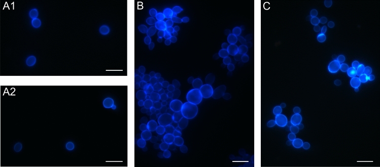
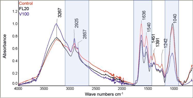
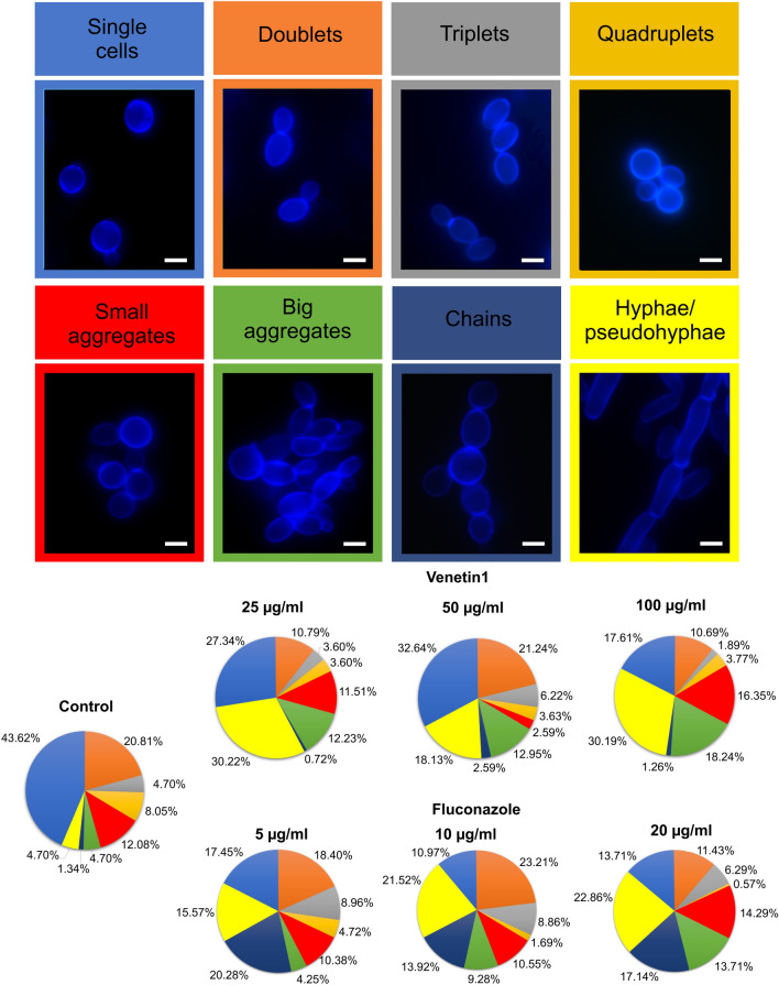
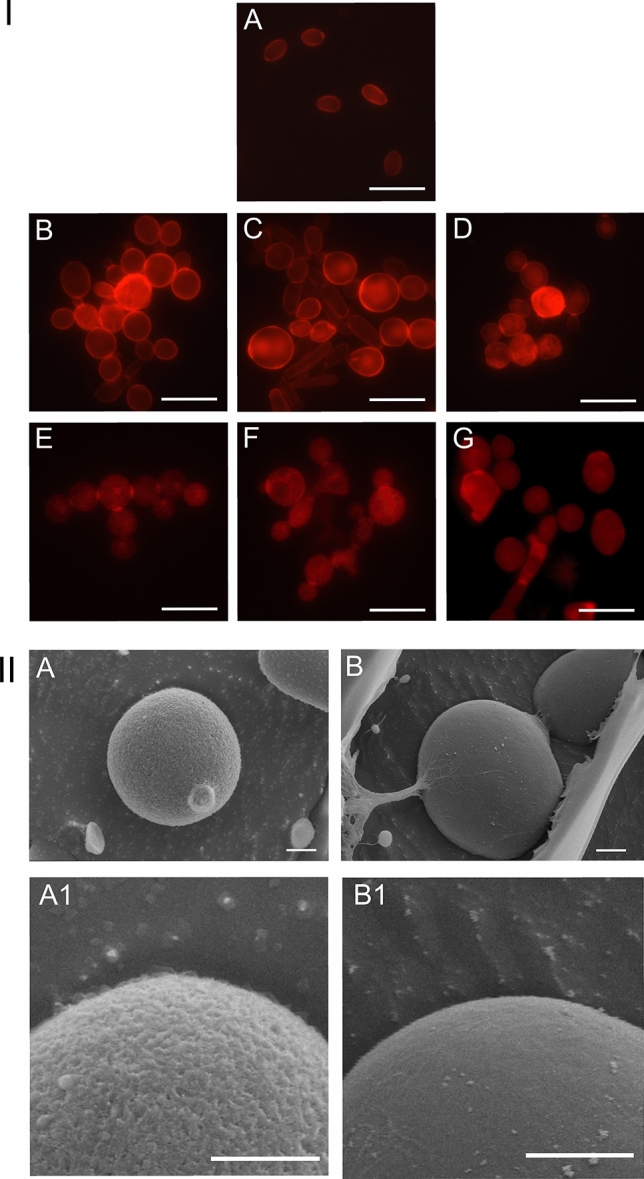
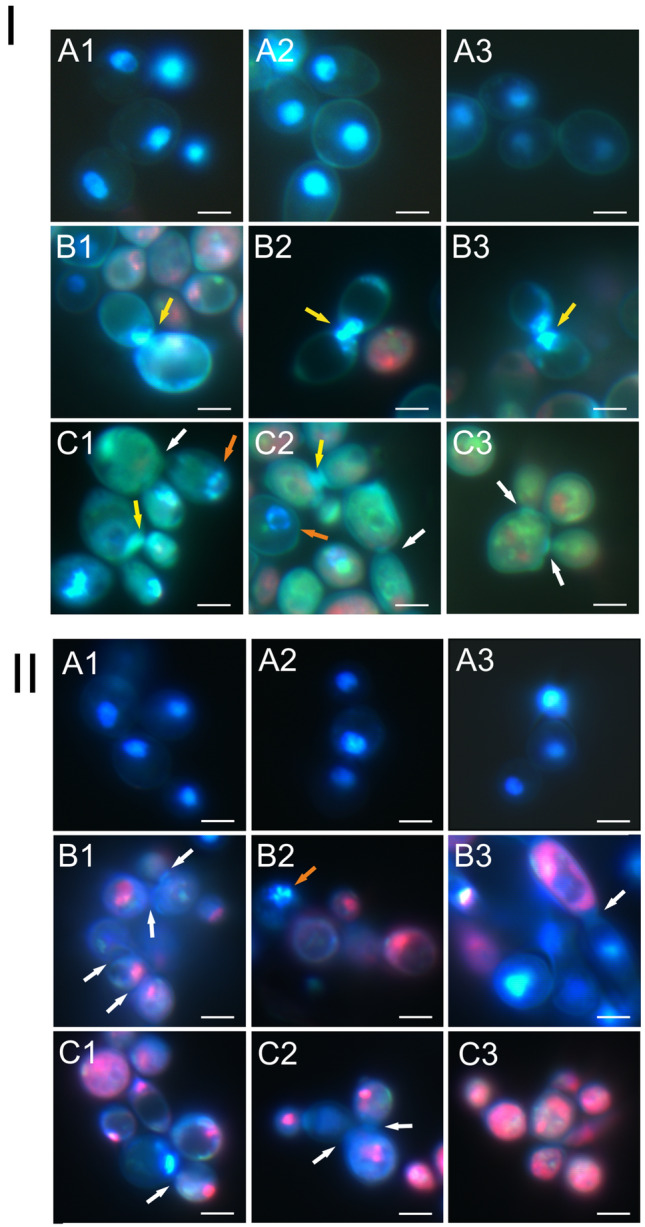
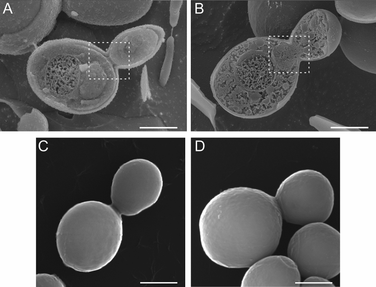
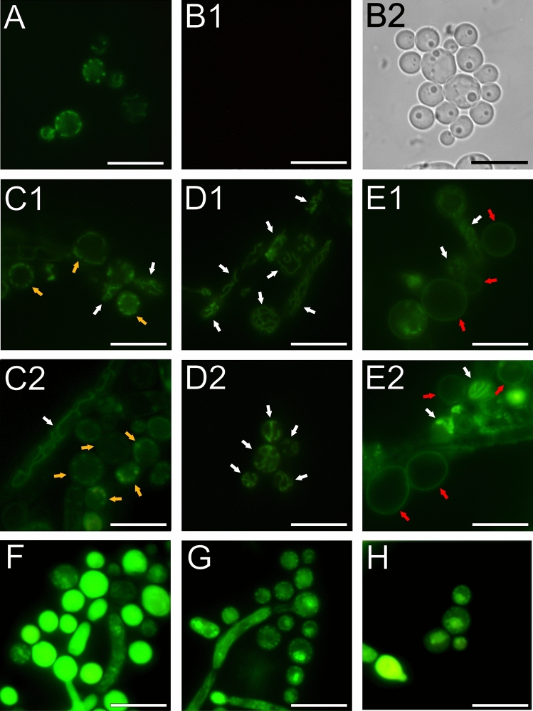
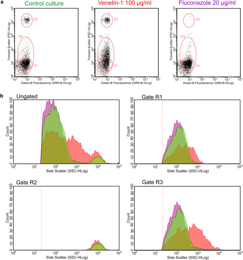
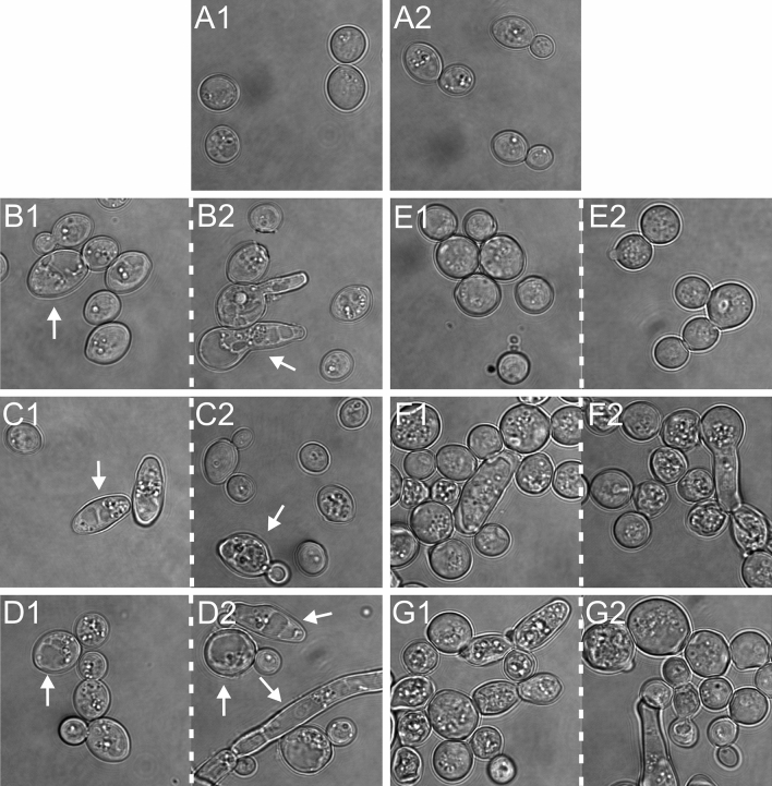
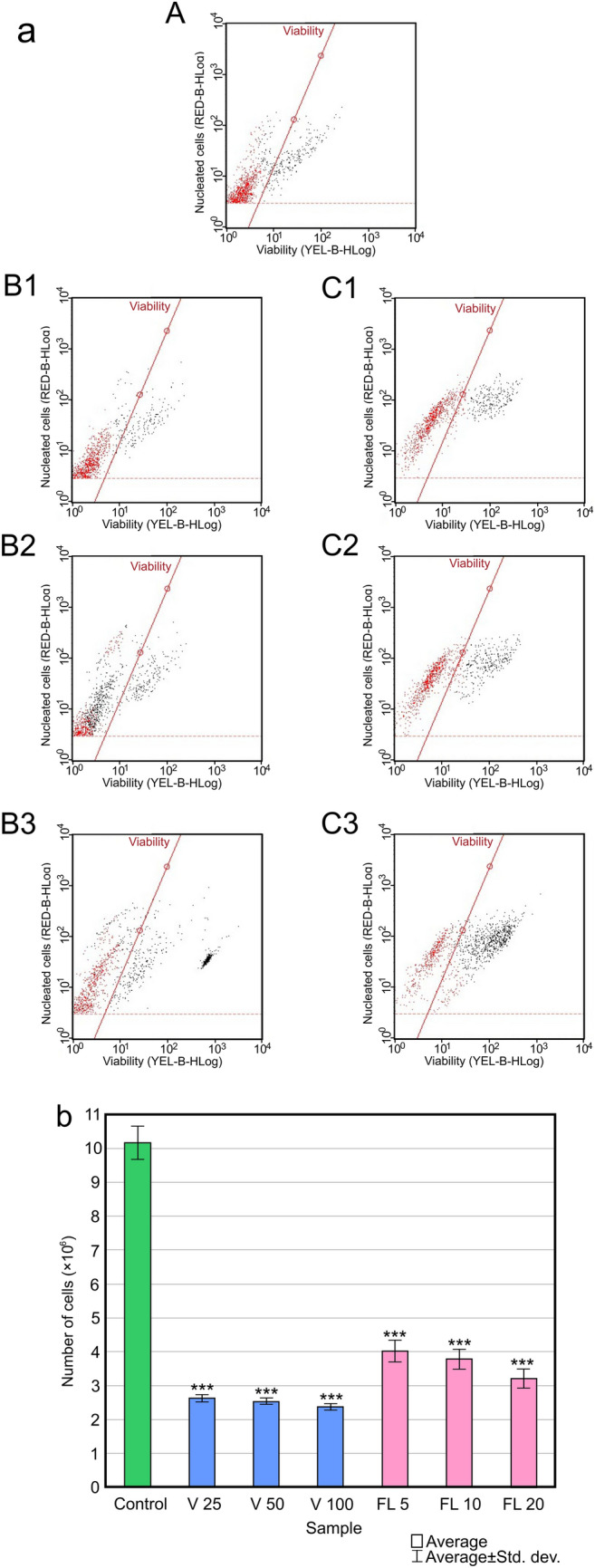
In the present research, the effect of a protein-polysaccharide complex Venetin-1 obtained from the coelomic fluid of Dendrobaena veneta earthworm on Candida albicans cells was characterized. The compound destroyed fungal cells without showing cytotoxicity to human skin fibroblasts, which was demonstrated in earlier studies. Since it had an effect on the fungal cell wall and membrane, this complex was compared with the known antifungal antibiotic fluconazole. Both preparations disturbed the division of yeast cells and resulted in the formation of aggregates and chains of unseparated cells, which was illustrated by staining with fluorochromes. Fluorescent staining of the cell wall with Calcofluor white facilitated comparison of the types of aggregates formed after the action of both substances. The analysis performed with the use of Congo red showed that Venetin-1 exposed deeper layers of the cell wall, whereas no such effect was visible after the use of fluconazole. The FTIR analysis confirmed changes in the mannoprotein layer of the cell wall after the application of the Venetin-1 complex. Staining with Rhodamine 123 and the use of flow cytometry allowed comparison of changes in the mitochondria. Significantly elongated mitochondria were observed after the Venetin-1 application, but not after the application of the classic antibiotic. Phase contrast microscopy revealed vacuole enlargement after the Venetin-1 application. The flow cytometry analysis of C. albicans cells treated with Venetin-1 and fluconazole showed that both substances caused a significant decrease in cell viability.
在本研究中,表征了从赤子爱胜蚓体腔液中获得的蛋白质-多糖复合物 Venetin-1 对白色念珠菌细胞的影响。该化合物在先前的研究中证明对人类皮肤成纤维细胞无细胞毒性,但能破坏真菌细胞。由于它对真菌细胞壁和膜有影响,因此将该复合物与已知的抗真菌抗生素氟康唑进行了比较。两种制剂都干扰了酵母细胞的分裂,导致未分离细胞的聚集和链状形成,这通过荧光染料染色得到了说明。用 Calcofluor white 对细胞壁进行荧光染色,便于比较两种物质作用后形成的聚集类型。用刚果红进行的分析表明,Venetin-1 暴露了细胞壁的更深层,而氟康唑则没有这种效果。FTIR 分析证实了细胞壁甘露糖蛋白层在应用 Venetin-1 复合物后的变化。用 Rhodamine 123 染色和流式细胞术的使用允许比较线粒体的变化。在用 Venetin-1 处理后观察到线粒体明显伸长,但在用经典抗生素处理后则没有。相差显微镜显示应用 Venetin-1 后液泡增大。用 Venetin-1 和氟康唑处理的 C. albicans 细胞的流式细胞术分析表明,两种物质都导致细胞活力显著下降。