Roh Hyeonhee, Otgondemberel Yanjinsuren, Eom Jeonghyeon, Kim Daniel, Im Maesoon
Brain Science Institute, Korea Institute of Science and Technology, Seoul, Republic of Korea.
School of Electrical Engineering, Korea University, Seoul, Republic of Korea.
Front Cell Neurosci. 2023 Feb 6;17:1115703. doi: 10.3389/fncel.2023.1115703. eCollection 2023.
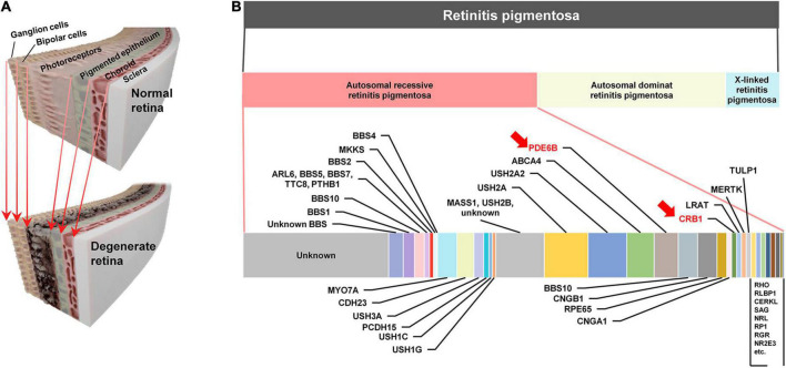
Microelectronic prostheses for artificial vision stimulate neurons surviving outer retinal neurodegeneration such as retinitis pigmentosa (RP). Yet, the quality of prosthetic vision substantially varies across subjects, maybe due to different levels of retinal degeneration and/or distinct genotypes. Although the RP genotypes are remarkably diverse, prosthetic studies have primarily used retinal degeneration () 1 and 10 mice, which both have gene mutation. Here, we report the electric responses arising in retinal ganglion cells (RGCs) of the mouse model which has mutation.
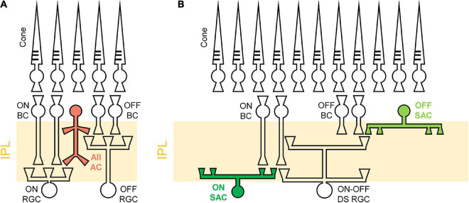
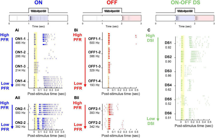
We first investigated age-dependent histological changes of wild-type (), , and mice retinas by H&E staining. Then, we used cell-attached patch clamping to record spiking responses of ON, OFF and direction selective (DS) types of RGCs to a 4-ms-long electric pulse. The electric responses of RGCs were analyzed in comparison with those of RGCs in terms of individual RGC spiking patterns, populational characteristics, and spiking consistency across trials.
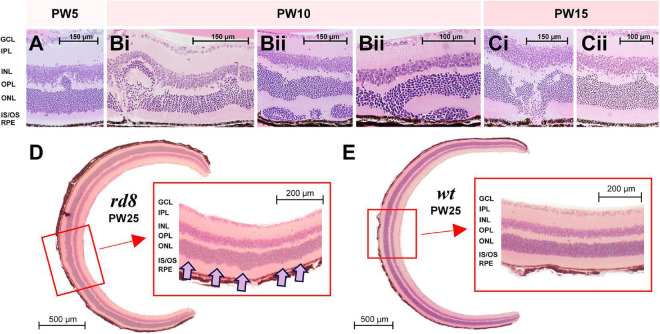
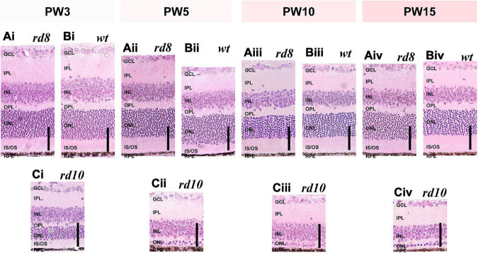
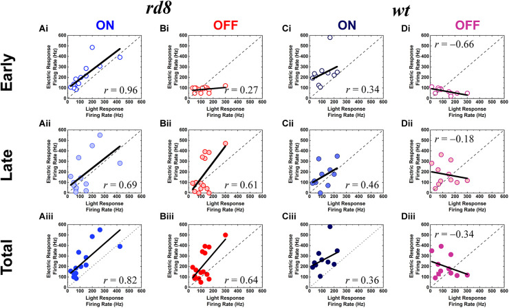
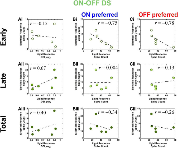
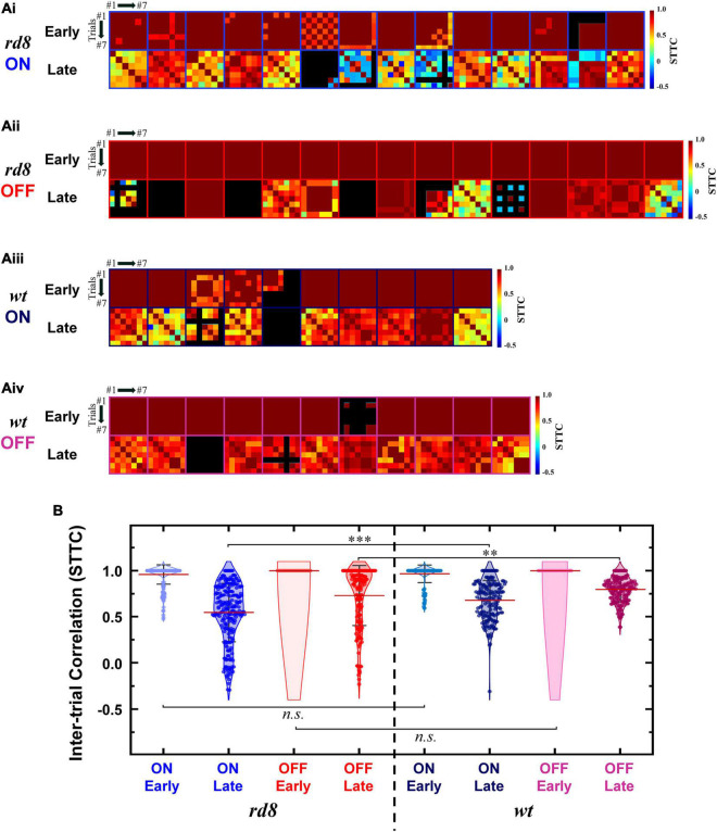
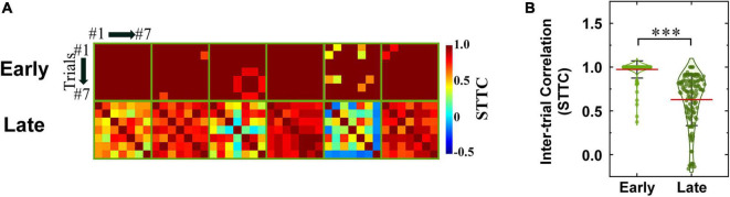
In the histological examination, the mice showed partial retinal foldings, but the outer nuclear layer thicknesses remained comparable to those of the mice, indicating the early-stage of RP. Although spiking patterns of each RGC type seemed similar to those of the retinas, correlation levels between electric vs. light response features were different across the two mouse models. For example, in comparisons between light vs. electric response magnitudes, ON/OFF RGCs of the mice showed the same/opposite correlation polarity with those of mice, respectively. Also, the electric response spike counts of DS RGCs in the retinas showed a positive correlation with their direction selectivity indices ( = 0.40), while those of the retinas were negatively correlated ( = -0.90). Lastly, the spiking timing consistencies of late responses were largely decreased in both ON and OFF RGCs in the than the retinas, whereas no significant difference was found across DS RGCs of the two models.
Our results indicate the electric response features are altered depending on RGC types even from the early-stage RP caused by mutation. Given the various degeneration patterns depending on mutation genes, our study suggests the importance of both genotype- and RGC type-dependent analyses for retinal prosthetic research.
用于人工视觉的微电子假体刺激诸如视网膜色素变性(RP)等外层视网膜神经退行性病变中存活的神经元。然而,假体视觉的质量在不同受试者之间有很大差异,这可能是由于视网膜变性程度不同和/或基因型不同所致。尽管RP基因型种类繁多,但假体研究主要使用视网膜变性(rd)1和10小鼠,这两种小鼠都有Pde6b基因突变。在此,我们报告了具有Pde6brd10突变的小鼠模型视网膜神经节细胞(RGC)产生的电反应。
我们首先通过苏木精-伊红(H&E)染色研究野生型(WT)、rd1和rd10小鼠视网膜的年龄依赖性组织学变化。然后,我们使用细胞贴附式膜片钳记录ON、OFF和方向选择性(DS)类型的RGC对4毫秒长电脉冲的放电反应。将rd10 RGC的电反应与rd1 RGC的电反应在单个RGC放电模式、群体特征以及不同试验间放电一致性方面进行比较分析。
在组织学检查中,rd10小鼠表现出部分视网膜折叠,但外核层厚度与rd1小鼠的相当,表明处于RP早期阶段。虽然每种RGC类型的放电模式似乎与rd1视网膜的相似,但两种小鼠模型中电反应与光反应特征之间的相关水平不同。例如,在比较光反应与电反应幅度时,rd10小鼠的ON/OFF RGC分别与rd1小鼠的呈现相同/相反的相关极性。此外,rd10视网膜中DS RGC的电反应尖峰计数与其方向选择性指数呈正相关(r = 0.40),而rd1视网膜的则呈负相关(r = -0.90)。最后,与rd1视网膜相比,rd10视网膜中ON和OFF RGC晚期反应的放电时间一致性在很大程度上降低,而两种模型的DS RGC之间未发现显著差异。
我们的结果表明,即使是由Pde6brd10突变引起的RP早期阶段,电反应特征也会因RGC类型而异。鉴于取决于突变基因的各种变性模式,我们的研究表明在视网膜假体研究中进行基因型和RGC类型依赖性分析的重要性。