Sobhani Navid, Bouchè Victoria, Aldegheri Giovanni, Rocca Andrea, D'Angelo Alberto, Giudici Fabiola, Bottin Cristina, Donofrio Carmine Antonio, Pinamonti Maurizio, Ferrari Benvenuto, Panni Stefano, Cominetti Marika, Aliaga Jahard, Ungari Marco, Fioravanti Antonio, Zanconati Fabrizio, Generali Daniele
Department of Medicine, Section of Epidemiology and Population Sciences, Baylor College of Medicine, Houston, TX 77030, USA.
Department of Medical, Surgery and Health Sciences, University of Trieste, 34147 Trieste, Italy.
Biomedicines. 2023 Jan 22;11(2):311. doi: 10.3390/biomedicines11020311.
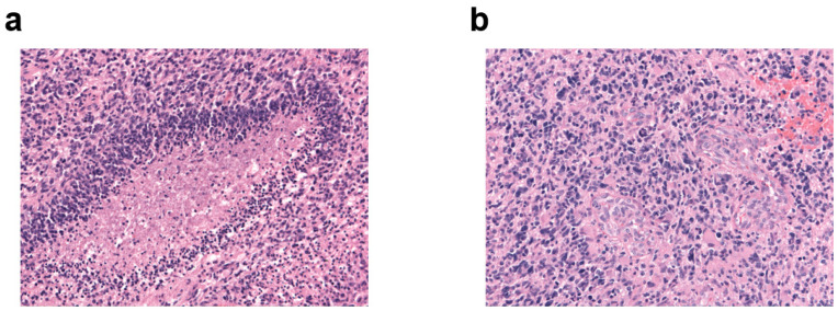
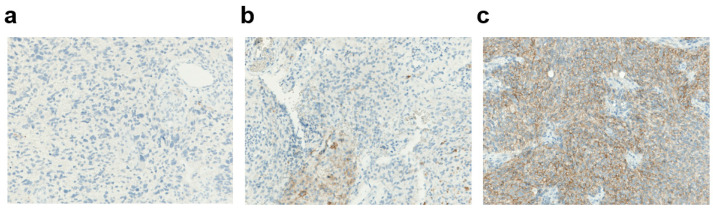
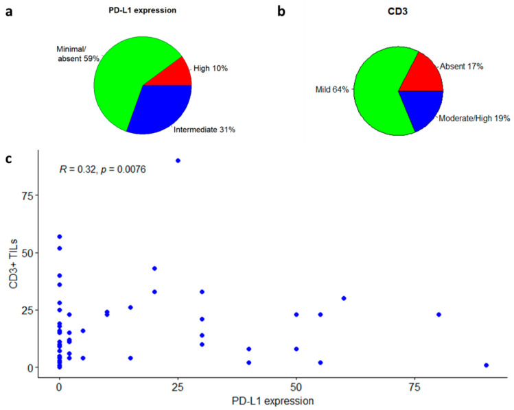
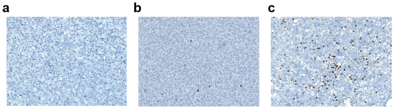
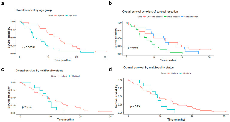
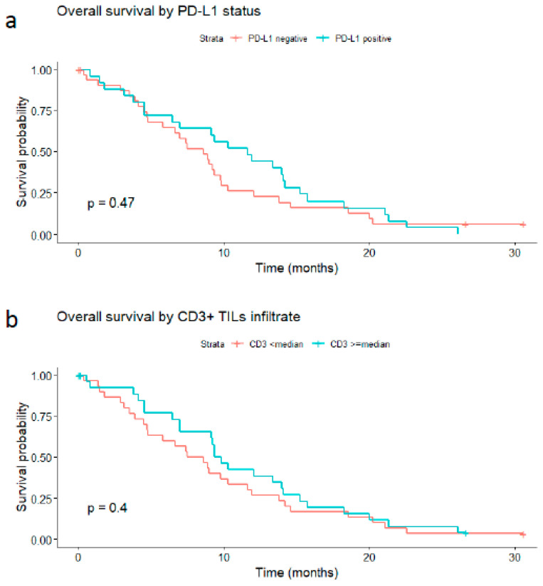
With the advent of immunotherapies, the field of cancer therapy has been revived with new hope, especially for cancers with dismal prognoses, such as the glioblastoma multiforme (GBM). Currently, immunotherapies should potentiate the host's own antitumor immune response against cancer cells, but it has been documented that they are effective only in small subsets of patients. Therefore, accurate predictors of response are urgently needed to identify who will benefit from immune-modulatory therapies. Brain tumors are challenging in terms of treatments. The immune response in the brain is highly regulated, and the immune microenvironment in brain metastases is active with a high density of tumor-infiltrating lymphocytes (TILs, CD3+ T cells) in certain patients and, therefore, may serve as a potential treatment target. In our study, we performed immunohistochemistry for CD3 and PD-L1 along the routine assessment of the O6-methylguanine-methyltransferase (MGMT) promoter methylation status and the IDH1 and 2 status in a single center cohort of 69 patients with GBM (58 primary tumors and 11 recurrences) who underwent standard multimodal therapies (surgery/radiotherapy/adjuvant temozolamide). We analyzed the association of PD-L1 tumor expression and TILs with overall survival (OS). The PD-L1 expression was observed in 25 of 58 (43%) newly diagnosed primary glioblastoma specimens. The sparse-to-moderate density of TILs, identified with CD3+ expression, was found in 48 of 58 (83%) specimens. Neither PD-L1 expression nor TILs were associated with overall survival. In conclusion, TILs and/or PD-L1 expression are detectable in the majority of glioblastoma samples, and even if they slightly relate to the outcome, they do not show a statistically significant correlation.
随着免疫疗法的出现,癌症治疗领域迎来了新的希望,尤其是对于预后不佳的癌症,如多形性胶质母细胞瘤(GBM)。目前,免疫疗法应增强宿主自身针对癌细胞的抗肿瘤免疫反应,但据记载,它们仅在一小部分患者中有效。因此,迫切需要准确的反应预测指标来确定谁将从免疫调节疗法中获益。脑肿瘤在治疗方面具有挑战性。大脑中的免疫反应受到高度调节,并且在某些患者中,脑转移瘤中的免疫微环境活跃,肿瘤浸润淋巴细胞(TILs,CD3 + T细胞)密度高,因此可能作为潜在的治疗靶点。在我们的研究中,我们在一个由69例接受标准多模式治疗(手术/放疗/辅助替莫唑胺)的GBM患者(58例原发性肿瘤和11例复发肿瘤)的单中心队列中,在常规评估O6 - 甲基鸟嘌呤 - 甲基转移酶(MGMT)启动子甲基化状态以及异柠檬酸脱氢酶1和2状态的同时,对CD3和PD - L1进行了免疫组织化学检测。我们分析了PD - L1肿瘤表达和TILs与总生存期(OS)的相关性。在58例新诊断的原发性胶质母细胞瘤标本中的25例(43%)中观察到了PD - L1表达。在58例标本中的48例(83%)中发现了以CD3 +表达确定的稀疏至中等密度的TILs。PD - L1表达和TILs均与总生存期无关。总之,在大多数胶质母细胞瘤样本中可检测到TILs和/或PD - L1表达,即使它们与结果略有相关,但未显示出统计学上的显著相关性。