Maralescu Felix-Mihai, Vaduva Adrian, Schiller Adalbert, Petrica Ligia, Sporea Ioan, Popescu Alina, Sirli Roxana, Dema Alis, Bodea Madalina, Grosu Iulia, Bob Flaviu
Division of Nephrology, Department of Internal Medicine II, Victor Babeș University of Medicine and Pharmacy, 300041 Timisoara, Romania.
Centre for Molecular Research in Nephrology and Vascular Disease, Victor Babeș University of Medicine and Pharmacy, 300041 Timisoara, Romania.
Biomedicines. 2023 Jan 26;11(2):365. doi: 10.3390/biomedicines11020365.
A renal biopsy represents the gold standard in the diagnosis, prognosis, and management of patients with glomerulonephritis. So far, non-invasive elastographic techniques have not confirmed their utility in replacing a biopsy; however, the new and improved software from Hologic Supersonic Mach 30 is a promising method for assessing the renal tissue's stiffness and viscosity. We investigated whether this elastography technique could reveal renal tissue fibrosis in patients with chronic glomerulonephritis.
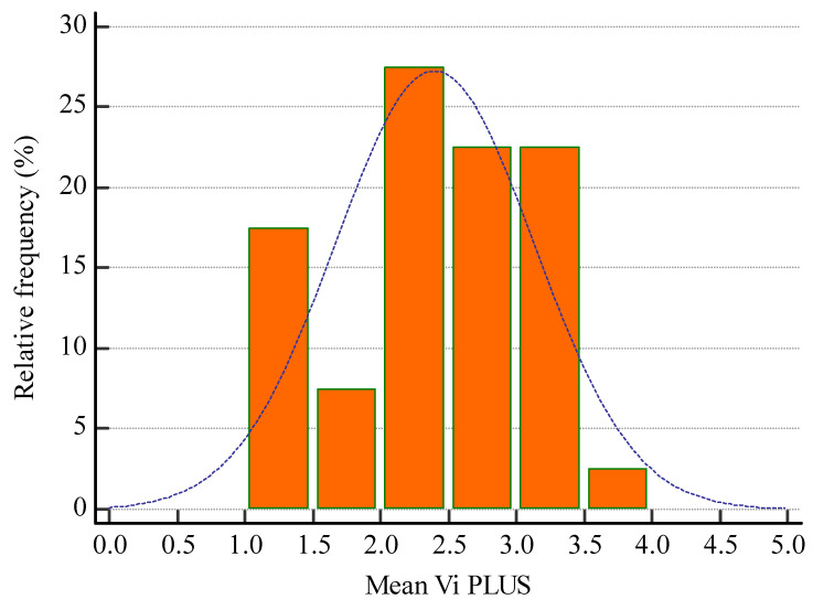
Two-dimensional-shear wave elastography (SWE) PLUS and viscosity plane-wave ultrasound (Vi PLUS) assessments were performed in 40 patients with chronic glomerulopathies before being referred for a renal biopsy. For each kidney, the mean values of five stiffness and viscosity measures were compared with the demographic, biological, and histopathological parameters of the patients.
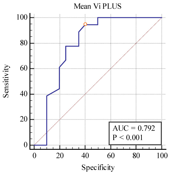
In total, 26 men and 14 women with a mean age of 52.35 ± 15.54 years, a mean estimated glomerular filtration rate (eGFR) of 53.8 ± 35.49 mL/min/1.73m, and a mean proteinuria of 6.39 ± 7.42 g/24 h were included after providing their informed consent. Out of 40 kidney biopsies, 2 were uninterpretable with inappropriate material and were divided into four subgroups based on their fibrosis percentage. Even though these elastography techniques were unable to differentiate between separate fibrosis stages, when predicting between the fibrosis and no-fibrosis group, we found a cut-off value of <20.77 kPa with the area under the curve (AUC) of 0.860, a < 0.001 with 88.89% sensitivity, and a 75% specificity for the 2D SWE PLUS measures and a cut-off value of <2.8 Pa.s with an AUC of 0.792, a < 0.001 with 94% sensitivity, and a 60% specificity for the Vi PLUS measures. We also found a cut-off value of <19.75 kPa for the 2D SWE PLUS measures (with an AUC of 0.789, = 0.0001 with 100% sensitivity, and a 74.29% specificity) and a cut-off value of <1.28 Pa.s for the Vi PLUS measures (with an AUC 0.829, = 0.0019 with 60% sensitivity, and a 94.29% specificity) differentiating between patients with over 40% fibrosis and those with under 40%. We also discovered a positive correlation between the glomerular filtration rate (eGFR) and 2D-SWE PLUS values ( = 0.7065, < 0.0001) and Vi PLUS values ( = 0.3637, < 0.0211). C reactive protein (CRP) correlates with the Vi PLUS measures (r = -0.3695, = 0.0189) but not with the 2D SWE PLUS measures ( = -0.2431, = 0.1306).
Our findings indicate that this novel elastography method can distinguish between individuals with different stages of renal fibrosis, correlate with the renal function and inflammation, and are easy to use and reproducible, but further research is needed for them to be employed routinely in clinical practice.
肾活检是肾小球肾炎患者诊断、预后评估及治疗的金标准。到目前为止,非侵入性弹性成像技术尚未证实其可替代活检的效用;然而,Hologic Supersonic Mach 30的新型改进软件是评估肾组织硬度和黏度的一种有前景的方法。我们研究了这种弹性成像技术能否揭示慢性肾小球肾炎患者的肾组织纤维化情况。
在40例慢性肾小球疾病患者进行肾活检前,采用二维剪切波弹性成像(SWE)PLUS和黏度平面波超声(Vi PLUS)进行评估。对于每个肾脏,将五个硬度和黏度测量值的平均值与患者的人口统计学、生物学和组织病理学参数进行比较。
共有26名男性和14名女性参与,平均年龄为52.35±15.54岁,平均估计肾小球滤过率(eGFR)为53.8±35.49 mL/min/1.73m²,平均蛋白尿为6.39±7.42 g/24 h,所有患者均签署了知情同意书。在40例肾活检中,2例因取材不当无法解读,其余病例根据纤维化百分比分为四个亚组。尽管这些弹性成像技术无法区分不同的纤维化阶段,但在预测纤维化组和无纤维化组时,我们发现二维SWE PLUS测量的截断值<20.77 kPa,曲线下面积(AUC)为0.860,P<0.001,灵敏度为88.89%,特异性为75%;Vi PLUS测量的截断值<2.8 Pa·s,AUC为0.792,P<0.001,灵敏度为94%,特异性为60%。我们还发现,二维SWE PLUS测量区分纤维化超过40%和低于40%患者的截断值<19.75 kPa(AUC为0.789,P = 0.0001,灵敏度为100%,特异性为74.29%),Vi PLUS测量的截断值<1.28 Pa·s(AUC为0.829,P = 0.0019,灵敏度为60%,特异性为94.29%)。我们还发现肾小球滤过率(eGFR)与二维SWE PLUS值(r = 0.7065,P<0.0001)和Vi PLUS值(r = 0.3637,P<0.021)呈正相关。C反应蛋白(CRP)与Vi PLUS测量值相关(r = -0.3695,P = 0.0189),但与二维SWE PLUS测量值无关(r = -0.2431,P = 0.1306)。
我们的研究结果表明,这种新型弹性成像方法能够区分不同阶段的肾纤维化个体,与肾功能和炎症相关,且易于使用和重复,但在临床实践中常规应用还需要进一步研究。