• 文献检索
  • 文档翻译
  • 深度研究
  • 学术资讯
  • Suppr Zotero 插件Zotero 插件
  • 邀请有礼
  • 套餐&价格
  • 历史记录
应用&插件
Suppr Zotero 插件Zotero 插件浏览器插件Mac 客户端Windows 客户端微信小程序
定价
高级版会员购买积分包购买API积分包
服务
文献检索文档翻译深度研究API 文档MCP 服务
关于我们
关于 Suppr公司介绍联系我们用户协议隐私条款
关注我们

Suppr 超能文献

核心技术专利:CN118964589B侵权必究
粤ICP备2023148730 号-1Suppr @ 2026

文献检索

告别复杂PubMed语法,用中文像聊天一样搜索,搜遍4000万医学文献。AI智能推荐,让科研检索更轻松。

立即免费搜索

文件翻译

保留排版,准确专业,支持PDF/Word/PPT等文件格式,支持 12+语言互译。

免费翻译文档

深度研究

AI帮你快速写综述,25分钟生成高质量综述,智能提取关键信息,辅助科研写作。

立即免费体验

缺氧条件下近端肾小管上皮细胞对足细胞损伤的旁分泌作用是由精氨酸酶-II 和 TGF-β1 介导的。

Paracrine Effects of Renal Proximal Tubular Epithelial Cells on Podocyte Injury under Hypoxic Conditions Are Mediated by Arginase-II and TGF-β1.

机构信息

Cardiovascular & Aging Research, Department of Endocrinology, Metabolism, Cardiovascular System, Faculty of Science and Medicine, University of Fribourg, CH-1700 Fribourg, Switzerland.

出版信息

Int J Mol Sci. 2023 Feb 10;24(4):3587. doi: 10.3390/ijms24043587.

DOI:10.3390/ijms24043587
PMID:36835007
原文链接:https://pmc.ncbi.nlm.nih.gov/articles/PMC9966309/
Abstract

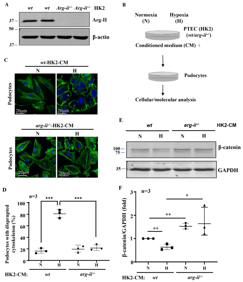
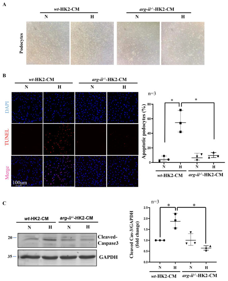
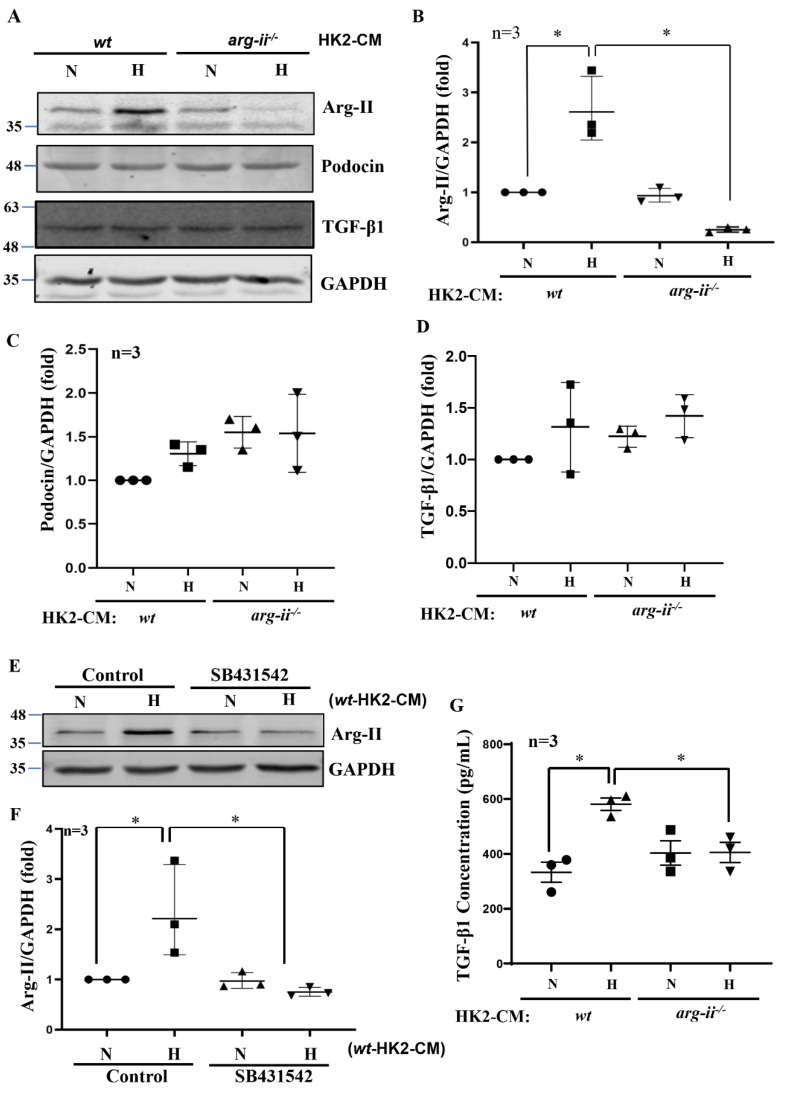
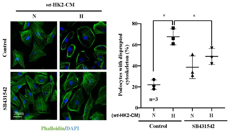
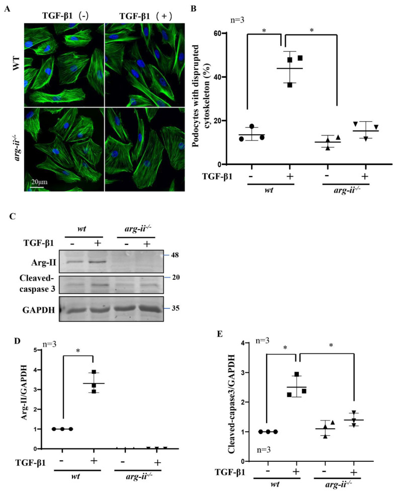
Hypoxia is an important risk for renal disease. The mitochondrial enzyme arginase-II (Arg-II) is expressed and/or induced by hypoxia in proximal tubular epithelial cells (PTECs) and in podocytes, leading to cellular damage. Because PTECs are vulnerable to hypoxia and located in proximity to podocytes, we examined the role of Arg-II in the crosstalk of PTECs under hypoxic conditions with podocytes. A human PTEC cell line (HK2) and a human podocyte cell line (AB8/13) were cultured. gene was ablated by CRISPR/Case9 in both cell types. HK2 cells were exposed to normoxia (21% O) or hypoxia (1% O) for 48 h. Conditioned medium (CM) was collected and transferred to the podocytes. Podocyte injuries were then analyzed. Hypoxic (not normoxic) HK2-CM caused cytoskeletal derangement, cell apoptosis, and increased Arg-II levels in differentiated podocytes. These effects were absent when in HK2 was ablated. The detrimental effects of the hypoxic HK2-CM were prevented by TGF-β1 type-I receptor blocker SB431542. Indeed, TGF-β1 levels in hypoxic HK2-CM (but not -HK2-CM) were increased. Furthermore, the detrimental effects of TGF-β1 on podocytes were prevented in -podocytes. This study demonstrates crosstalk between PTECs and podocytes through the Arg-II-TGF-β1 cascade, which may contribute to hypoxia-induced podocyte damage.

摘要

缺氧是肾脏疾病的一个重要危险因素。线粒体酶精氨酸酶-II(Arg-II)在近端肾小管上皮细胞(PTEC)和足细胞中由缺氧表达和/或诱导,导致细胞损伤。由于 PTEC 对缺氧敏感且位于足细胞附近,因此我们研究了 Arg-II 在缺氧条件下 PTEC 与足细胞相互作用中的作用。培养人 PTEC 细胞系(HK2)和人足细胞系(AB8/13)。在这两种细胞类型中,通过 CRISPR/Cas9 敲除 基因。将 HK2 细胞暴露于常氧(21% O)或缺氧(1% O)48 小时。收集条件培养基(CM)并转移至足细胞。然后分析足细胞损伤。缺氧(而非常氧)HK2-CM 导致分化的足细胞骨架紊乱、细胞凋亡和 Arg-II 水平升高。当在 HK2 中敲除 时,这些作用不存在。TGF-β1 型 I 型受体阻滞剂 SB431542 可预防缺氧 HK2-CM 的有害作用。事实上,缺氧 HK2-CM(而非 -HK2-CM)中的 TGF-β1 水平增加。此外,TGF-β1 对 -podocytes 的有害作用也被预防。这项研究表明,PTEC 和足细胞通过 Arg-II-TGF-β1 级联相互作用,这可能导致缺氧诱导的足细胞损伤。

https://cdn.ncbi.nlm.nih.gov/pmc/blobs/fbd4/9966309/ea9c747fcc4c/ijms-24-03587-g006.jpg
https://cdn.ncbi.nlm.nih.gov/pmc/blobs/fbd4/9966309/77a389d2010d/ijms-24-03587-g001.jpg
https://cdn.ncbi.nlm.nih.gov/pmc/blobs/fbd4/9966309/50456c694599/ijms-24-03587-g002.jpg
https://cdn.ncbi.nlm.nih.gov/pmc/blobs/fbd4/9966309/7323158cf2f4/ijms-24-03587-g003.jpg
https://cdn.ncbi.nlm.nih.gov/pmc/blobs/fbd4/9966309/9f160035ff27/ijms-24-03587-g004.jpg
https://cdn.ncbi.nlm.nih.gov/pmc/blobs/fbd4/9966309/071c9c94d160/ijms-24-03587-g005.jpg
https://cdn.ncbi.nlm.nih.gov/pmc/blobs/fbd4/9966309/ea9c747fcc4c/ijms-24-03587-g006.jpg

相似文献

1
Paracrine Effects of Renal Proximal Tubular Epithelial Cells on Podocyte Injury under Hypoxic Conditions Are Mediated by Arginase-II and TGF-β1.缺氧条件下近端肾小管上皮细胞对足细胞损伤的旁分泌作用是由精氨酸酶-II 和 TGF-β1 介导的。
Int J Mol Sci. 2023 Feb 10;24(4):3587. doi: 10.3390/ijms24043587.
2
Role of Arginase-II in Podocyte Injury under Hypoxic Conditions.缺氧条件下诱导型一氧化氮合酶在足细胞损伤中的作用。
Biomolecules. 2022 Aug 31;12(9):1213. doi: 10.3390/biom12091213.
3
TGF-β1 alters DNA methylation levels in promoter and enhancer regions of the WT1 gene in human podocytes.转化生长因子-β1改变人足细胞中WT1基因启动子和增强子区域的DNA甲基化水平。
Nephrology (Carlton). 2019 May;24(5):575-584. doi: 10.1111/nep.13411.
4
Response of Renal Podocytes to Excessive Hydrostatic Pressure: a Pathophysiologic Cascade in a Malignant Hypertension Model.肾小球足细胞对过高静水压的反应:恶性高血压模型中的病理生理级联反应。
Kidney Blood Press Res. 2017;42(6):1104-1118. doi: 10.1159/000485774. Epub 2017 Dec 7.
5
Hypoxia Induces Renal Epithelial Injury and Activates Fibrotic Signaling Through Up-Regulation of Arginase-II.缺氧通过上调精氨酸酶II诱导肾上皮损伤并激活纤维化信号通路。
Front Physiol. 2021 Nov 19;12:773719. doi: 10.3389/fphys.2021.773719. eCollection 2021.
6
Renal tubular Bim mediates the tubule-podocyte crosstalk via NFAT2 to induce podocyte cytoskeletal dysfunction.Bim 在肾小管中通过 NFAT2 介导管周细胞的串扰,导致足细胞细胞骨架功能障碍。
Theranostics. 2020 May 22;10(15):6806-6824. doi: 10.7150/thno.43145. eCollection 2020.
7
TGF-β1 stimulates movement of renal proximal tubular epithelial cells in a three-dimensional cell culture via an autocrine TGF-β2 production.转化生长因子-β1通过自分泌产生转化生长因子-β2,在三维细胞培养中刺激肾近端小管上皮细胞的迁移。
Exp Cell Res. 2017 Jan 1;350(1):132-139. doi: 10.1016/j.yexcr.2016.11.014. Epub 2016 Nov 21.
8
Role of pulmonary epithelial arginase-II in activation of fibroblasts and lung inflammaging.肺上皮细胞精氨酸酶-II 在成纤维细胞激活和肺炎症中的作用。
Aging Cell. 2023 Apr;22(4):e13790. doi: 10.1111/acel.13790. Epub 2023 Feb 15.
9
High glucose increases Cdk5 activity in podocytes via transforming growth factor-β1 signaling pathway.高葡萄糖通过转化生长因子-β1 信号通路增加足细胞中的 Cdk5 活性。
Exp Cell Res. 2014 Aug 15;326(2):219-29. doi: 10.1016/j.yexcr.2014.04.014. Epub 2014 Apr 24.
10
microRNA in Extracellular Vesicles Released by Damaged Podocytes Promote Apoptosis of Renal Tubular Epithelial Cells.损伤的足细胞释放的细胞外囊泡中的 microRNA 促进肾小管上皮细胞凋亡。
Cells. 2020 Jun 5;9(6):1409. doi: 10.3390/cells9061409.

引用本文的文献

1
Crosstalk between glomeruli and tubules.肾小球与肾小管之间的相互作用。
Nat Rev Nephrol. 2025 Mar;21(3):189-199. doi: 10.1038/s41581-024-00907-0. Epub 2024 Dec 6.
2
Elevation of Arginase-II in Podocytes Contributes to Age-Associated Albuminuria in Male Mice.足细胞中精氨酸酶-II 的升高导致雄性小鼠与年龄相关的白蛋白尿。
Int J Mol Sci. 2023 Jul 7;24(13):11228. doi: 10.3390/ijms241311228.

本文引用的文献

1
Role of Arginase-II in Podocyte Injury under Hypoxic Conditions.缺氧条件下诱导型一氧化氮合酶在足细胞损伤中的作用。
Biomolecules. 2022 Aug 31;12(9):1213. doi: 10.3390/biom12091213.
2
Prescribing SGLT2 Inhibitors in Patients With CKD: Expanding Indications and Practical Considerations.为慢性肾脏病患者开具钠-葡萄糖协同转运蛋白2抑制剂:适应证拓展与实际考量
Kidney Int Rep. 2022 May 5;7(7):1463-1476. doi: 10.1016/j.ekir.2022.04.094. eCollection 2022 Jul.
3
Mechanistic Pathogenesis of Endothelial Dysfunction in Diabetic Nephropathy and Retinopathy.
糖尿病肾病和视网膜病变中内皮功能障碍的发病机制。
Front Endocrinol (Lausanne). 2022 May 25;13:816400. doi: 10.3389/fendo.2022.816400. eCollection 2022.
4
Tubular cells produce FGF2 via autophagy after acute kidney injury leading to fibroblast activation and renal fibrosis.急性肾损伤后,管状细胞通过自噬产生 FGF2,导致成纤维细胞活化和肾纤维化。
Autophagy. 2023 Jan;19(1):256-277. doi: 10.1080/15548627.2022.2072054. Epub 2022 May 18.
5
Mechanisms of podocyte injury and implications for diabetic nephropathy.足细胞损伤的机制及其在糖尿病肾病中的意义。
Clin Sci (Lond). 2022 Apr 14;136(7):493-520. doi: 10.1042/CS20210625.
6
Hypoxia and chronic kidney disease.缺氧与慢性肾脏病。
EBioMedicine. 2022 Mar;77:103942. doi: 10.1016/j.ebiom.2022.103942. Epub 2022 Mar 13.
7
The Proximal Tubule as the Pathogenic and Therapeutic Target in Acute Kidney Injury.近端肾小管作为急性肾损伤的致病和治疗靶点。
Nephron. 2022;146(5):494-502. doi: 10.1159/000522341. Epub 2022 Mar 10.
8
The multifaced role of HtrA1 in the development of joint and skeletal disorders.HtrA1在关节和骨骼疾病发展中的多方面作用。
Bone. 2022 Apr;157:116350. doi: 10.1016/j.bone.2022.116350. Epub 2022 Feb 5.
9
Hypoxia Induces Renal Epithelial Injury and Activates Fibrotic Signaling Through Up-Regulation of Arginase-II.缺氧通过上调精氨酸酶II诱导肾上皮损伤并激活纤维化信号通路。
Front Physiol. 2021 Nov 19;12:773719. doi: 10.3389/fphys.2021.773719. eCollection 2021.
10
PGC1α is required for the renoprotective effect of lncRNA Tug1 in vivo and links Tug1 with urea cycle metabolites.PGC1α 对于 lncRNA Tug1 在体内的肾保护作用是必需的,并将 Tug1 与尿素循环代谢物联系起来。
Cell Rep. 2021 Aug 10;36(6):109510. doi: 10.1016/j.celrep.2021.109510.