• 文献检索
  • 文档翻译
  • 深度研究
  • 学术资讯
  • Suppr Zotero 插件Zotero 插件
  • 邀请有礼
  • 套餐&价格
  • 历史记录
应用&插件
Suppr Zotero 插件Zotero 插件浏览器插件Mac 客户端Windows 客户端微信小程序
定价
高级版会员购买积分包购买API积分包
服务
文献检索文档翻译深度研究API 文档MCP 服务
关于我们
关于 Suppr公司介绍联系我们用户协议隐私条款
关注我们

Suppr 超能文献

核心技术专利:CN118964589B侵权必究
粤ICP备2023148730 号-1Suppr @ 2026

文献检索

告别复杂PubMed语法,用中文像聊天一样搜索,搜遍4000万医学文献。AI智能推荐,让科研检索更轻松。

立即免费搜索

文件翻译

保留排版,准确专业,支持PDF/Word/PPT等文件格式,支持 12+语言互译。

免费翻译文档

深度研究

AI帮你快速写综述,25分钟生成高质量综述,智能提取关键信息,辅助科研写作。

立即免费体验

Stefin B 淀粉样纤维呈现各向异性性质。

Amyloid Fibrils of Stefin B Show Anisotropic Properties.

机构信息

Department of Biochemistry and Molecular and Structural Biology, Jožef Stefan Institute, Jamova 39, 1000 Ljubljana, Slovenia.

Faculty of Mathematics and Physics, University of Ljubljana, Jadranska 19, 1000 Ljubljana, Slovenia.

出版信息

Int J Mol Sci. 2023 Feb 13;24(4):3737. doi: 10.3390/ijms24043737.

DOI:10.3390/ijms24043737
PMID:36835149
原文链接:https://pmc.ncbi.nlm.nih.gov/articles/PMC9962164/
Abstract

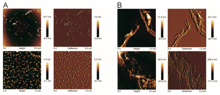
Human stefin B, a member of the cystatin family of cysteine protease inhibitors, tends to form amyloid fibrils under relatively mild conditions, which is why it is used as a model protein to study amyloid fibrillation. Here, we show for the first time that bundles of amyloid fibrils, i.e., helically twisted ribbons, formed by human stefin B exhibit birefringence. This physical property is commonly observed in amyloid fibrils when stained with Congo red. However, we show that the fibrils arrange in regular anisotropic arrays and no staining is required. They share this property with anisotropic protein crystals, structured protein arrays such as tubulin and myosin, and other anisotropic elongated materials, such as textile fibres and liquid crystals. In certain macroscopic arrangements of amyloid fibrils, not only birefringence is observed, but also enhanced emission of intrinsic fluorescence, implying a possibility to detect amyloid fibrils with no labels by using optical microscopy. In our case, no enhancement of intrinsic tyrosine fluorescence was observed at 303 nm; instead, an additional fluorescence emission peak appeared at 425 to 430 nm. We believe that both phenomena, birefringence and fluorescence emission in the deep blue, should be further explored with this and other amyloidogenic proteins. This may allow the development of label-free detection methods for amyloid fibrils of different origins.

摘要

人源组织蛋白酶抑制剂 B(Stefin B)属于半胱氨酸蛋白酶抑制剂家族成员,在相对温和的条件下易于形成淀粉样纤维,因此常被用作研究淀粉样纤维形成的模型蛋白。在此,我们首次证明由人源组织蛋白酶抑制剂 B 形成的淀粉样纤维束(即螺旋扭曲的带状物)具有双折射性。这一物理性质在使用刚果红染色的淀粉样纤维中很常见。然而,我们发现纤维呈规则各向异性排列,无需染色。它们与各向异性蛋白晶体、微管蛋白和肌球蛋白等结构化蛋白阵列以及其他各向异性细长材料(如纺织纤维和液晶)具有相同的性质。在某些淀粉样纤维的宏观排列中,不仅观察到双折射,还观察到固有荧光的增强发射,这意味着可以通过光学显微镜检测无标记的淀粉样纤维。在我们的情况下,在 303nm 处未观察到酪氨酸固有荧光的增强;相反,在 425 到 430nm 处出现了一个额外的荧光发射峰。我们相信,双折射和深蓝光下的荧光发射这两种现象都应该用这种和其他淀粉样蛋白进一步探索。这可能会为不同来源的淀粉样纤维开发无标记检测方法。

https://cdn.ncbi.nlm.nih.gov/pmc/blobs/d2ec/9962164/75aa076a9b01/ijms-24-03737-g004.jpg
https://cdn.ncbi.nlm.nih.gov/pmc/blobs/d2ec/9962164/7a58678bb679/ijms-24-03737-g001.jpg
https://cdn.ncbi.nlm.nih.gov/pmc/blobs/d2ec/9962164/24064ad7c565/ijms-24-03737-g002.jpg
https://cdn.ncbi.nlm.nih.gov/pmc/blobs/d2ec/9962164/2072837320a0/ijms-24-03737-g003.jpg
https://cdn.ncbi.nlm.nih.gov/pmc/blobs/d2ec/9962164/75aa076a9b01/ijms-24-03737-g004.jpg

相似文献

1
Amyloid Fibrils of Stefin B Show Anisotropic Properties.Stefin B 淀粉样纤维呈现各向异性性质。
Int J Mol Sci. 2023 Feb 13;24(4):3737. doi: 10.3390/ijms24043737.
2
Human stefin B readily forms amyloid fibrils in vitro.人源stefin B在体外易于形成淀粉样纤维。
Biochim Biophys Acta. 2002 Jan 31;1594(1):1-5. doi: 10.1016/s0167-4838(01)00295-3.
3
Different propensity to form amyloid fibrils by two homologous proteins-Human stefins A and B: searching for an explanation.两种同源蛋白——人抑制蛋白A和B形成淀粉样纤维的不同倾向:探寻一种解释。
Proteins. 2004 May 1;55(2):417-25. doi: 10.1002/prot.20041.
4
Amyloid fibril formation by human stefin B in vitro: immunogold labelling and comparison to stefin A.人stefin B在体外形成淀粉样纤维:免疫金标记及与stefin A的比较
Biol Chem. 2002 May;383(5):859-63. doi: 10.1515/BC.2002.092.
5
High affinity copper binding by stefin B (cystatin B) and its role in the inhibition of amyloid fibrillation.斯替芬B(胱抑素B)对铜的高亲和力结合及其在抑制淀粉样蛋白原纤维形成中的作用。
FEBS J. 2006 Sep;273(18):4250-63. doi: 10.1111/j.1742-4658.2006.05426.x.
6
Folding and amyloid-fibril formation for a series of human stefins' chimeras: any correlation?一系列人微小泛素相关修饰蛋白嵌合体的折叠与淀粉样纤维形成:有何关联?
Proteins. 2006 Mar 1;62(4):918-27. doi: 10.1002/prot.20812.
7
Amyloid fibril formation by human stefin B: influence of pH and TFE on fibril growth and morphology.人stefin B形成淀粉样纤维:pH值和三氟乙醇对纤维生长及形态的影响
Amyloid. 2007 Sep;14(3):237-47. doi: 10.1080/13506120701461137.
8
Physical basis of colors seen in Congo red-stained amyloid in polarized light.刚果红染色淀粉样蛋白在偏振光下呈现颜色的物理基础。
Lab Invest. 2008 Mar;88(3):232-42. doi: 10.1038/labinvest.3700714. Epub 2007 Dec 31.
9
Conformational changes preceding amyloid-fibril formation of amyloid-beta and stefin B; parallels in pH dependence.淀粉样β蛋白和丝抑蛋白B形成淀粉样纤维之前的构象变化;pH依赖性的相似之处
Curr Med Chem. 2002 Oct;9(19):1717-24. doi: 10.2174/0929867023369097.
10
Interaction of human stefin B in the prefibrillar oligomeric form with membranes. Correlation with cellular toxicity.人源前纤维寡聚体形式的丝氨酸蛋白酶抑制剂B与膜的相互作用。与细胞毒性的相关性。
FEBS J. 2005 Jun;272(12):3042-51. doi: 10.1111/j.1742-4658.2005.04717.x.

本文引用的文献

1
General Principles Underpinning Amyloid Structure.淀粉样蛋白结构的基本原理。
Front Neurosci. 2022 Jun 2;16:878869. doi: 10.3389/fnins.2022.878869. eCollection 2022.
2
Role of aromatic amino acids in amyloid self-assembly.芳香族氨基酸在淀粉样蛋白自组装中的作用。
Int J Biol Macromol. 2020 Aug 1;156:949-959. doi: 10.1016/j.ijbiomac.2020.03.064. Epub 2020 Mar 19.
3
Six-fold director field configuration in amyloid nematic and cholesteric phases.六重 director 场在向列相和胆甾相中的组态。
Sci Rep. 2019 Sep 2;9(1):12654. doi: 10.1038/s41598-019-48996-3.
4
Exploring the potential of deep-blue autofluorescence for monitoring amyloid fibril formation and dissociation.探索深蓝色自发荧光在监测淀粉样纤维形成和解离方面的潜力。
PeerJ. 2019 Aug 16;7:e7554. doi: 10.7717/peerj.7554. eCollection 2019.
5
Dissection of the deep-blue autofluorescence changes accompanying amyloid fibrillation.解析伴随淀粉样纤维形成的深蓝色自发荧光变化。
Arch Biochem Biophys. 2018 Aug 1;651:13-20. doi: 10.1016/j.abb.2018.05.019. Epub 2018 May 24.
6
Carbonyl-based blue autofluorescence of proteins and amino acids.蛋白质和氨基酸基于羰基的蓝色自发荧光。
PLoS One. 2017 May 25;12(5):e0176983. doi: 10.1371/journal.pone.0176983. eCollection 2017.
7
Label-free imaging of amyloids using their intrinsic linear and nonlinear optical properties.利用淀粉样蛋白固有的线性和非线性光学特性对其进行无标记成像。
Biomed Opt Express. 2017 Jan 10;8(2):743-756. doi: 10.1364/BOE.8.000743. eCollection 2017 Feb 1.
8
Amyloid fibrils compared to peptide nanotubes.
Biochim Biophys Acta. 2014 Sep;1840(9):2944-52. doi: 10.1016/j.bbagen.2014.05.019. Epub 2014 Jun 5.
9
Structural and optical properties of short peptides: nanotubes-to-nanofibers phase transformation.短肽的结构与光学性质:从纳米管到纳米纤维的相变
J Pept Sci. 2014 Jul;20(7):487-93. doi: 10.1002/psc.2661. Epub 2014 Jun 3.
10
The role of initial oligomers in amyloid fibril formation by human stefin B.人源组织蛋白酶 B 形成淀粉样纤维过程中初始寡聚物的作用
Int J Mol Sci. 2013 Sep 5;14(9):18362-84. doi: 10.3390/ijms140918362.