Centre for 3D Models of Health and Disease, Department of Targeted Intervention, Division of Surgery and Interventional Science, University College London, Charles Bell House, 43-45 Foley Street, London W1W 7TS, UK.
Department of Mechanical Engineering, University College London, Gower Street, London WC1E 6BT, UK.
Int J Mol Sci. 2023 Feb 16;24(4):3956. doi: 10.3390/ijms24043956.
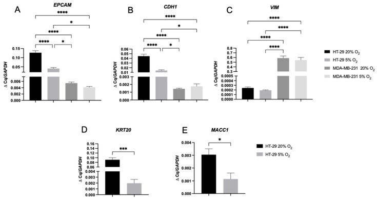
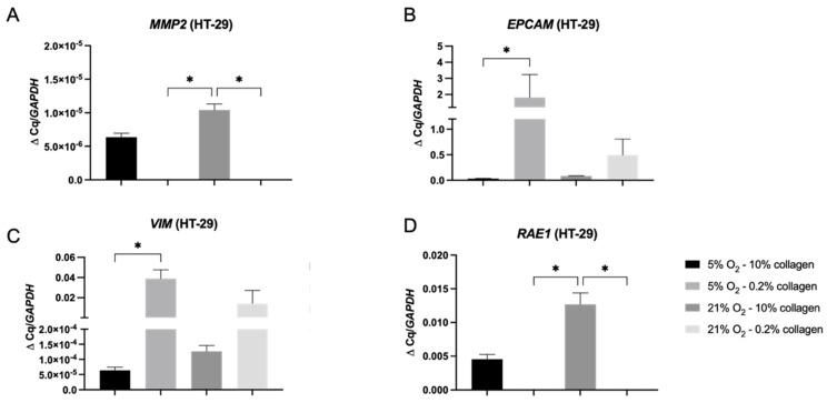
Epithelial to mesenchymal transition (EMT) in cancer is the process described where cancer epithelial cells acquire mesenchymal properties which can lead to enhanced invasiveness. Three-dimensional cancer models often lack the relevant and biomimetic microenvironment parameters appropriate to the native tumour microenvironment thought to drive EMT. In this study, HT-29 epithelial colorectal cells were cultivated in different oxygen and collagen concentrations to investigate how these biophysical parameters influenced invasion patterns and EMT. Colorectal HT-29 cells were grown in physiological hypoxia (5% O) and normoxia (21% O) in 2D, 3D soft (60 Pa), and 3D stiff (4 kPa) collagen matrices. Physiological hypoxia was sufficient to trigger expression of markers of EMT in the HT-29 cells in 2D by day 7. This is in contrast to a control breast cancer cell line, MDA-MB-231, which expresses a mesenchymal phenotype regardless of the oxygen concentration. In 3D, HT-29 cells invaded more extensively in a stiff matrix environment with corresponding increases in the invasive genes and . This demonstrates that the physiological environment can directly impact HT-29 cells in terms of EMT marker expression and invasion, compared to an established cell line, MDA-MB-231, which has already undergone EMT. This study highlights the importance of the biophysical microenvironment to cancer epithelial cells and how these factors can direct cell behaviour. In particular, that stiffness of the 3D matrix drives greater invasion in HT-29 cells regardless of hypoxia. It is also pertinent that some cell lines (already having undergone EMT) are not as sensitive to the biophysical features of their microenvironment.
上皮间质转化(EMT)在癌症中是指描述癌症上皮细胞获得间充质特性的过程,这可能导致侵袭性增强。三维癌症模型通常缺乏适当的相关仿生微环境参数,这些参数被认为是驱动 EMT 的原始肿瘤微环境的一部分。在这项研究中,HT-29 上皮结肠癌细胞在不同的氧和胶原蛋白浓度下培养,以研究这些生物物理参数如何影响侵袭模式和 EMT。结肠 HT-29 细胞在生理缺氧(5% O)和常氧(21% O)下在 2D、3D 软(60 Pa)和 3D 硬(4 kPa)胶原蛋白基质中生长。生理缺氧足以在第 7 天在 2D 中触发 HT-29 细胞 EMT 标志物的表达。这与对照乳腺癌细胞系 MDA-MB-231 形成对比,后者无论氧浓度如何都表达间充质表型。在 3D 中,HT-29 细胞在刚性基质环境中更广泛地侵袭,相应地增加了侵袭基因 和 。这表明,与已经经历 EMT 的 MDA-MB-231 等已建立的细胞系相比,生理环境可以直接影响 HT-29 细胞的 EMT 标志物表达和侵袭。本研究强调了生物物理微环境对癌症上皮细胞的重要性,以及这些因素如何指导细胞行为。特别是 3D 基质的刚性无论缺氧如何都能驱动 HT-29 细胞的更大侵袭。同样重要的是,一些细胞系(已经经历 EMT)对其微环境的生物物理特征不那么敏感。