Pesapane Filippo, De Marco Paolo, Rapino Anna, Lombardo Eleonora, Nicosia Luca, Tantrige Priyan, Rotili Anna, Bozzini Anna Carla, Penco Silvia, Dominelli Valeria, Trentin Chiara, Ferrari Federica, Farina Mariagiorgia, Meneghetti Lorenza, Latronico Antuono, Abbate Francesca, Origgi Daniela, Carrafiello Gianpaolo, Cassano Enrico
Breast Imaging Division, IEO European Institute of Oncology IRCCS, 20141 Milan, Italy.
Medical Physics Unit, IEO European Institute of Oncology IRCCS, 20141 Milan, Italy.
J Clin Med. 2023 Feb 9;12(4):1372. doi: 10.3390/jcm12041372.
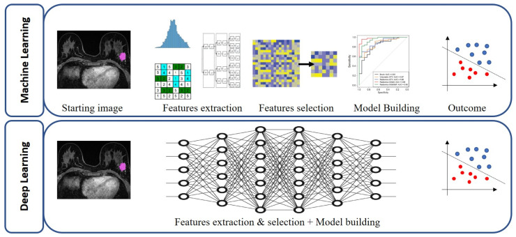
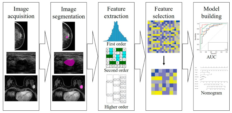
Recent technological advances in the field of artificial intelligence hold promise in addressing medical challenges in breast cancer care, such as early diagnosis, cancer subtype determination and molecular profiling, prediction of lymph node metastases, and prognostication of treatment response and probability of recurrence. Radiomics is a quantitative approach to medical imaging, which aims to enhance the existing data available to clinicians by means of advanced mathematical analysis using artificial intelligence. Various published studies from different fields in imaging have highlighted the potential of radiomics to enhance clinical decision making. In this review, we describe the evolution of AI in breast imaging and its frontiers, focusing on handcrafted and deep learning radiomics. We present a typical workflow of a radiomics analysis and a practical "how-to" guide. Finally, we summarize the methodology and implementation of radiomics in breast cancer, based on the most recent scientific literature to help researchers and clinicians gain fundamental knowledge of this emerging technology. Alongside this, we discuss the current limitations of radiomics and challenges of integration into clinical practice with conceptual consistency, data curation, technical reproducibility, adequate accuracy, and clinical translation. The incorporation of radiomics with clinical, histopathological, and genomic information will enable physicians to move forward to a higher level of personalized management of patients with breast cancer.
人工智能领域最近的技术进步有望应对乳腺癌护理中的医学挑战,如早期诊断、癌症亚型确定和分子特征分析、淋巴结转移预测以及治疗反应和复发概率的预后评估。放射组学是一种医学成像的定量方法,旨在通过使用人工智能的先进数学分析来增强临床医生可用的现有数据。来自不同成像领域的各种已发表研究突出了放射组学在改善临床决策方面的潜力。在这篇综述中,我们描述了人工智能在乳腺成像中的发展及其前沿领域,重点关注手工制作和深度学习放射组学。我们展示了放射组学分析的典型工作流程和实用的“操作方法”指南。最后,我们根据最新的科学文献总结了放射组学在乳腺癌中的方法和应用,以帮助研究人员和临床医生获得这一新兴技术的基础知识。与此同时,我们讨论了放射组学当前的局限性以及在概念一致性、数据管理、技术可重复性、足够的准确性和临床转化等方面融入临床实践的挑战。将放射组学与临床、组织病理学和基因组信息相结合,将使医生能够朝着更高水平的乳腺癌患者个性化管理迈进。