Zuhayri Hala, Samarinova Alice A, Borisov Alexey V, Guardado David A Lopez, Baalbaki Houssain, Krivova Natalya A, Kistenev Yury V
Laboratory of Laser Molecular Imaging and Machine Learning, Tomsk State University, Lenin Ave. 36, Tomsk 634050, Russia.
Pharmaceutics. 2023 Feb 10;15(2):595. doi: 10.3390/pharmaceutics15020595.
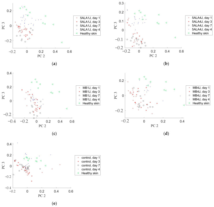
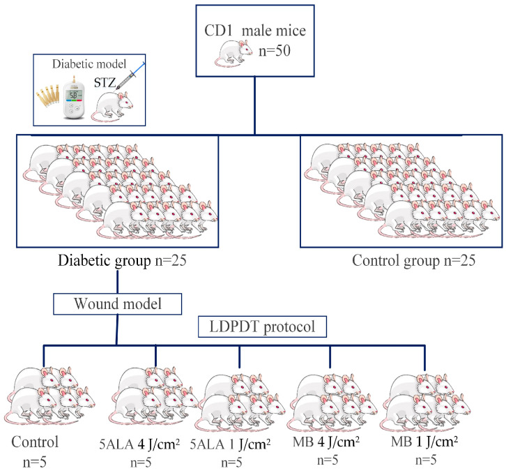
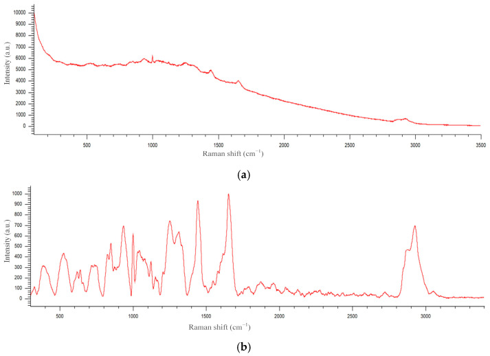
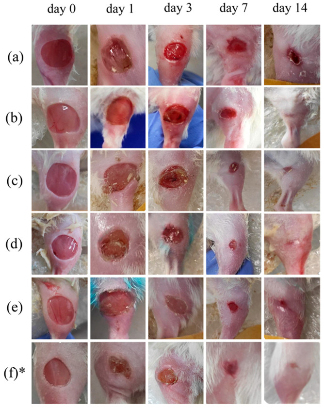
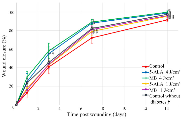
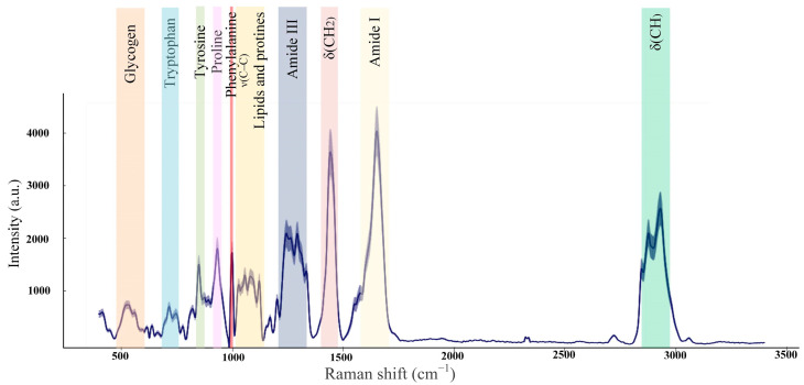
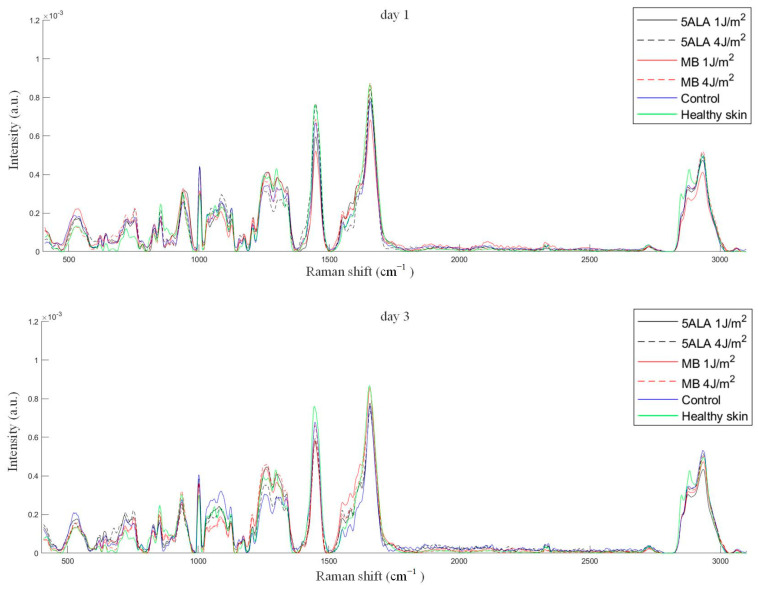
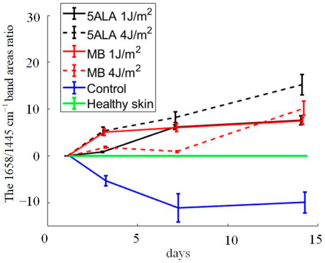
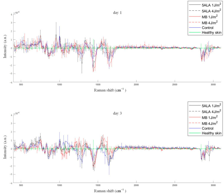
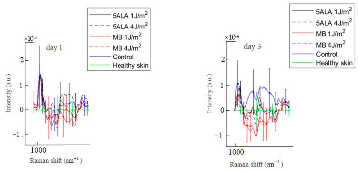
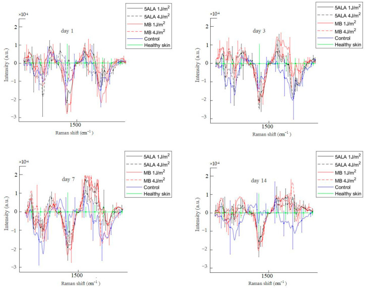
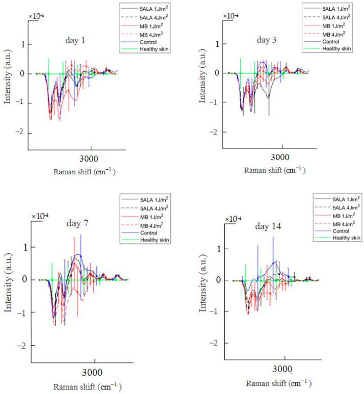
One of challenges that faces diabetes is the wound healing process. The delayed diabetic wound healing is caused by a complicated molecular mechanism involving numerous physiological variables. Low-dose photodynamic therapy (LDPDT) provides excellent results in rejuvenation and wound healing. In this study, the LDPDT effect on diabetic wounds in mice was studied using two photosensitizers, 5-aminolevulinic acid and methylene blue, and two laser dose expositions of 1 J/cm and 4 J/cm by Raman spectroscopy (RS). The latter was used as a noninvasive method, providing specific information about tissue state based on the fundamental vibrational modes of its molecular components. RS allows high spatial resolution acquisition of biochemical and structural information through the generation of point spectra or spectral images. An approach to in vivo quantitative assessment of diabetic wound healing state was developed. This approach is based on an application of the principal component analysis combined with the Mahalanobis metrics to skin Raman spectra, in particular, intensities of the amide I and CH bands.
糖尿病面临的挑战之一是伤口愈合过程。糖尿病伤口愈合延迟是由涉及众多生理变量的复杂分子机制引起的。低剂量光动力疗法(LDPDT)在皮肤修复和伤口愈合方面效果显著。在本研究中,通过拉曼光谱(RS),使用两种光敏剂(5-氨基酮戊酸和亚甲蓝)以及两种激光剂量照射(1 J/cm²和4 J/cm²),研究了LDPDT对小鼠糖尿病伤口的影响。后者作为一种非侵入性方法,基于其分子成分的基本振动模式提供有关组织状态的特定信息。RS通过生成点光谱或光谱图像,能够以高空间分辨率获取生化和结构信息。开发了一种体内定量评估糖尿病伤口愈合状态的方法。该方法基于将主成分分析与马氏距离度量应用于皮肤拉曼光谱,特别是酰胺I和CH波段的强度。