Fujii Shunsaku, Oguchi Takaaki
ef.clinic Aomori Japan.
Reprod Med Biol. 2023 Feb 23;22(1):e12508. doi: 10.1002/rmb2.12508. eCollection 2023 Jan-Dec.
A cross-sectional study was conducted to evaluate differences in uterine morphology between women with or without polycystic ovary syndrome.
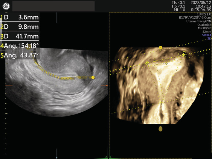
The authors recruited 333 infertile reproductive-age women including 93 with polycystic ovary syndrome diagnosed using the criteria of the Japanese Society of Obstetrics Gynecology-2007. Shapes of uterine cavity were measured by transvaginal three-dimensional ultrasound.
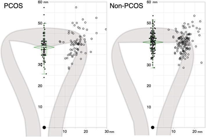
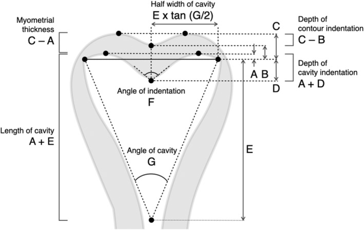
The polycystic ovary syndrome group had a significantly deeper indentation (2.2 ± 0.4 mm vs. 0.0 ± 0.2 mm, < 0.0001) and a significantly more acute indentation angle (162.9 ± 2.2 deg vs. 175.2 ± 1.3 deg, < 0.0001) than the control group.
The depth and the apical angle of fundal indentation of uterine cavity are different in women with polycystic ovary syndrome.
进行一项横断面研究,以评估患有或未患有多囊卵巢综合征的女性子宫形态的差异。
作者招募了333名育龄期不孕女性,其中93名根据日本妇产科学会2007年标准诊断为多囊卵巢综合征。通过经阴道三维超声测量子宫腔形态。
多囊卵巢综合征组的压痕明显更深(2.2±0.4毫米对0.0±0.2毫米,<0.0001),压痕角度明显更锐(162.9±2.2度对175.2±1.3度,<0.0001),均优于对照组。
多囊卵巢综合征女性子宫腔底部压痕的深度和顶端角度有所不同。