Chen Kaidong, Zhang Li, Mao Haixia, Chen Kefei, Shi Yachen, Meng Xiangpan, Wang Feng, Hu Xiaoyun, Fang Xiangming
Department of Radiology, The Affiliated Wuxi People's Hospital of Nanjing Medical University, Wuxi, China.
Department of Neurology, The Affiliated Wuxi People's Hospital of Nanjing Medical University, Wuxi, China.
Front Aging Neurosci. 2023 Feb 9;15:1116516. doi: 10.3389/fnagi.2023.1116516. eCollection 2023.
Anxiety is one of the most common psychiatric symptoms of Parkinson's disease (PD), and brain iron deposition is considered to be one of the pathological mechanisms of PD. The objective of this study was to explore alterations in brain iron deposition in PD patients with anxiety compared to PD patients without anxiety, especially in the fear circuit.
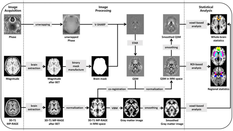
Sixteen PD patients with anxiety, 23 PD patients without anxiety, and 26 healthy elderly controls were enrolled prospectively. All subjects underwent neuropsychological assessments and brain magnetic resonance imaging (MRI) examinations. Voxel-based morphometry (VBM) was used to study morphological brain differences between the groups. Quantitative susceptibility mapping (QSM), an MRI technique capable of quantifying susceptibility changes in brain tissue, was used to compare susceptibility changes in the whole brain among the three groups. The correlations between brain susceptibility changes and anxiety scores quantified using the Hamilton Anxiety Rating Scale (HAMA) were compared and analyzed.
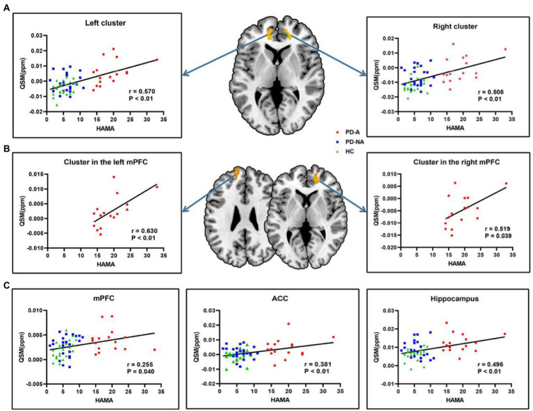
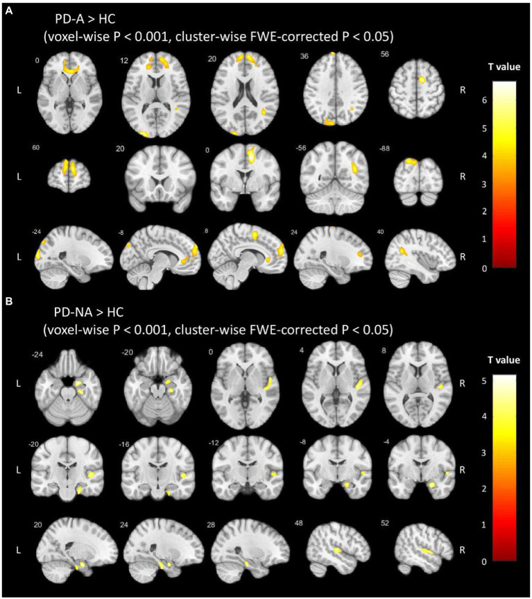
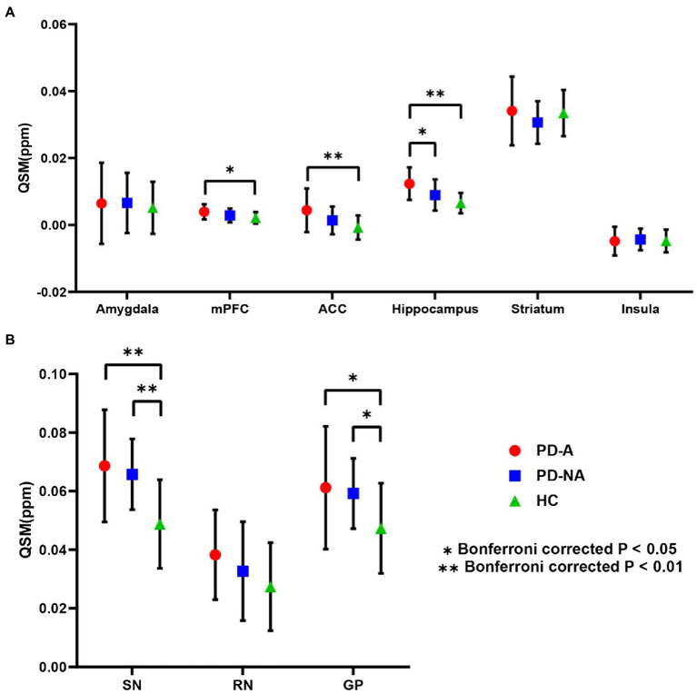
PD patients with anxiety had a longer duration of PD and higher HAMA scores than PD patients without anxiety. No morphological brain differences were observed between the groups. In contrast, voxel-based and ROI-based QSM analyses showed that PD patients with anxiety had significantly increased QSM values in the medial prefrontal cortex, anterior cingulate cortex, hippocampus, precuneus, and angular cortex. Furthermore, the QSM values of some of these brain regions were positively correlated with the HAMA scores (medial prefrontal cortex: = 0.255, = 0.04; anterior cingulate cortex: = 0.381, < 0.01; hippocampus: = 0.496, < 0.01).
Our findings support the idea that anxiety in PD is associated with iron burden in the brain fear circuit, providing a possible new approach to explaining the potential neural mechanism of anxiety in PD.
焦虑是帕金森病(PD)最常见的精神症状之一,脑铁沉积被认为是PD的病理机制之一。本研究的目的是探讨伴有焦虑的PD患者与不伴有焦虑的PD患者相比脑铁沉积的变化,尤其是在恐惧回路中的变化。
前瞻性纳入16例伴有焦虑的PD患者、23例不伴有焦虑的PD患者和26例健康老年对照者。所有受试者均接受神经心理学评估和脑磁共振成像(MRI)检查。基于体素的形态学测量(VBM)用于研究各组之间脑形态学差异。定量磁化率成像(QSM)是一种能够量化脑组织磁化率变化的MRI技术,用于比较三组全脑的磁化率变化。比较并分析使用汉密尔顿焦虑量表(HAMA)量化的脑磁化率变化与焦虑评分之间的相关性。
伴有焦虑的PD患者的PD病程长于不伴有焦虑的PD患者,且HAMA评分更高。各组之间未观察到脑形态学差异。相比之下,可以基于体素和感兴趣区域的QSM分析显示,伴有焦虑的PD患者在内侧前额叶皮质、前扣带回皮质、海马、楔前叶和角回皮质的QSM值显著增加。此外,这些脑区中的一些脑区的QSM值与HAMA评分呈正相关(内侧前额叶皮质:r = 0.255,P = 0.04;前扣带回皮质:r = 0.381,P < 0.01;海马:r = 0.496,P < 0.01)。
我们的研究结果支持以下观点,即PD中的焦虑与脑恐惧回路中的铁负荷有关,为解释PD中焦虑的潜在神经机制提供了一种可能的新方法。