• 文献检索
  • 文档翻译
  • 深度研究
  • 学术资讯
  • Suppr Zotero 插件Zotero 插件
  • 邀请有礼
  • 套餐&价格
  • 历史记录
应用&插件
Suppr Zotero 插件Zotero 插件浏览器插件Mac 客户端Windows 客户端微信小程序
定价
高级版会员购买积分包购买API积分包
服务
文献检索文档翻译深度研究API 文档MCP 服务
关于我们
关于 Suppr公司介绍联系我们用户协议隐私条款
关注我们

Suppr 超能文献

核心技术专利:CN118964589B侵权必究
粤ICP备2023148730 号-1Suppr @ 2026

文献检索

告别复杂PubMed语法,用中文像聊天一样搜索,搜遍4000万医学文献。AI智能推荐,让科研检索更轻松。

立即免费搜索

文件翻译

保留排版,准确专业,支持PDF/Word/PPT等文件格式,支持 12+语言互译。

免费翻译文档

深度研究

AI帮你快速写综述,25分钟生成高质量综述,智能提取关键信息,辅助科研写作。

立即免费体验

心肌梗死后的动态间质细胞反应可预测遗传多样性小鼠对破裂的抵抗力。

Dynamic Interstitial Cell Response during Myocardial Infarction Predicts Resilience to Rupture in Genetically Diverse Mice.

机构信息

The Jackson Laboratory, Bar Harbor, ME 04609, USA.

The Jackson Laboratory, Bar Harbor, ME 04609, USA.

出版信息

Cell Rep. 2020 Mar 3;30(9):3149-3163.e6. doi: 10.1016/j.celrep.2020.02.008.

DOI:10.1016/j.celrep.2020.02.008
PMID:32130914
原文链接:https://pmc.ncbi.nlm.nih.gov/articles/PMC7059115/
Abstract

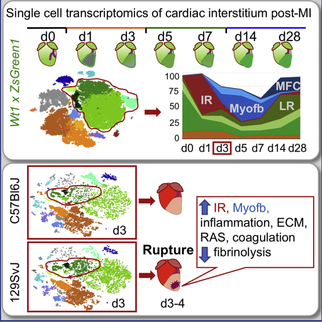
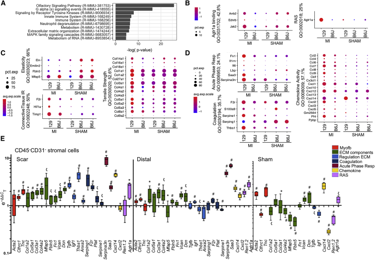
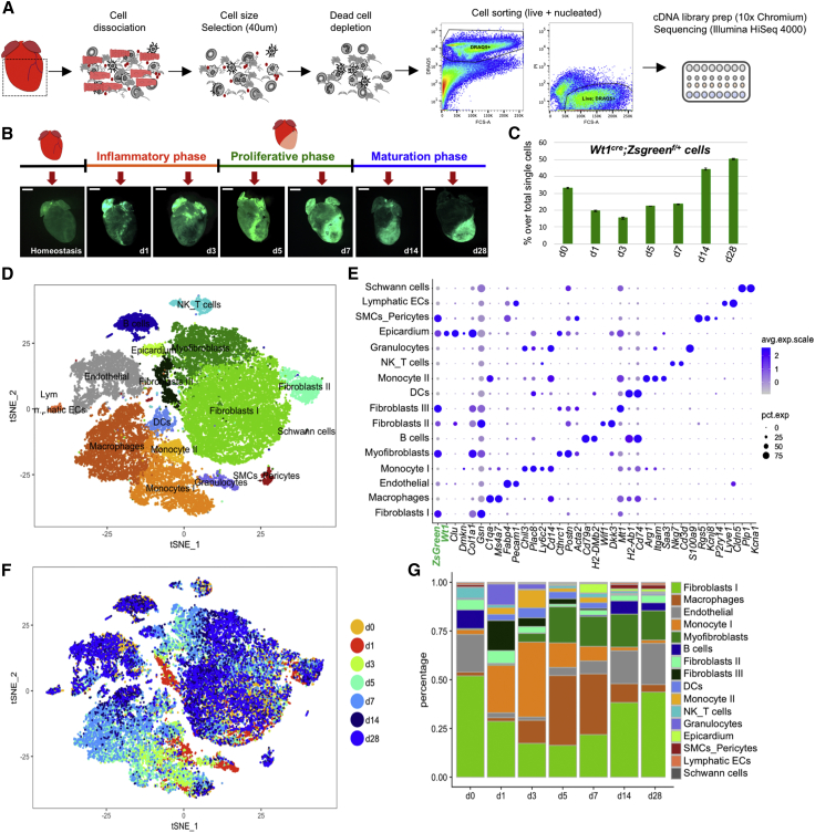
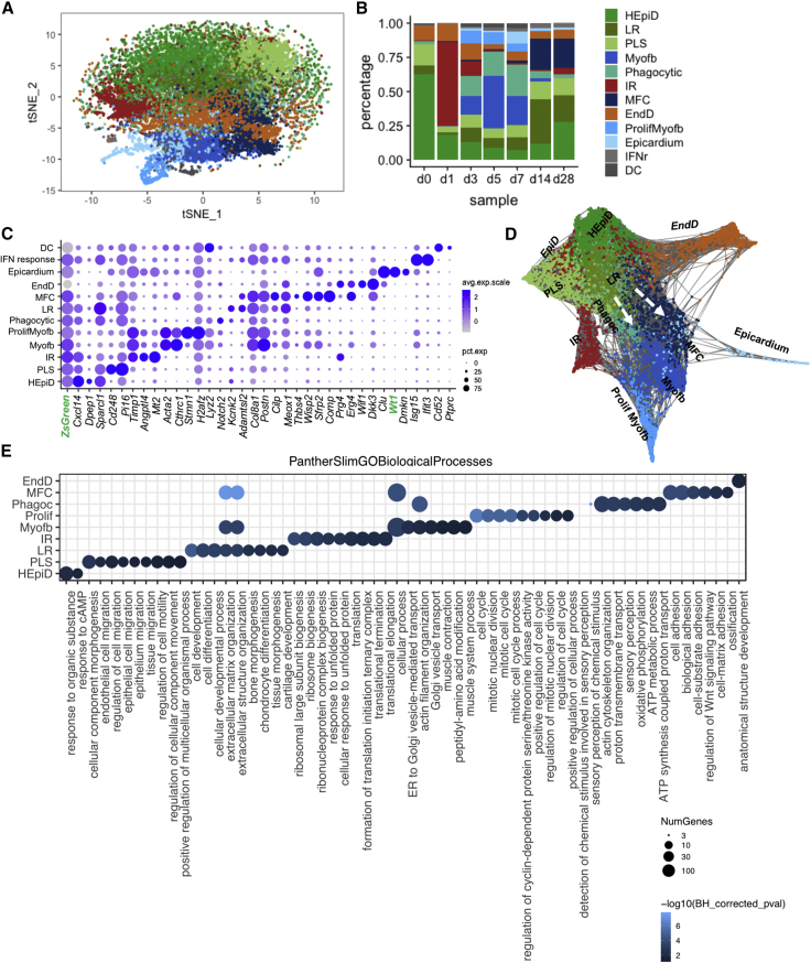
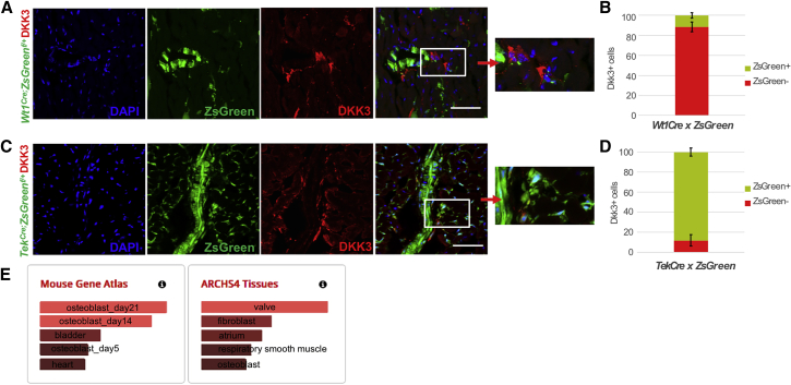
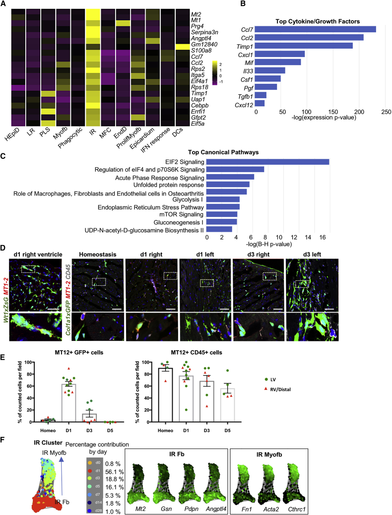
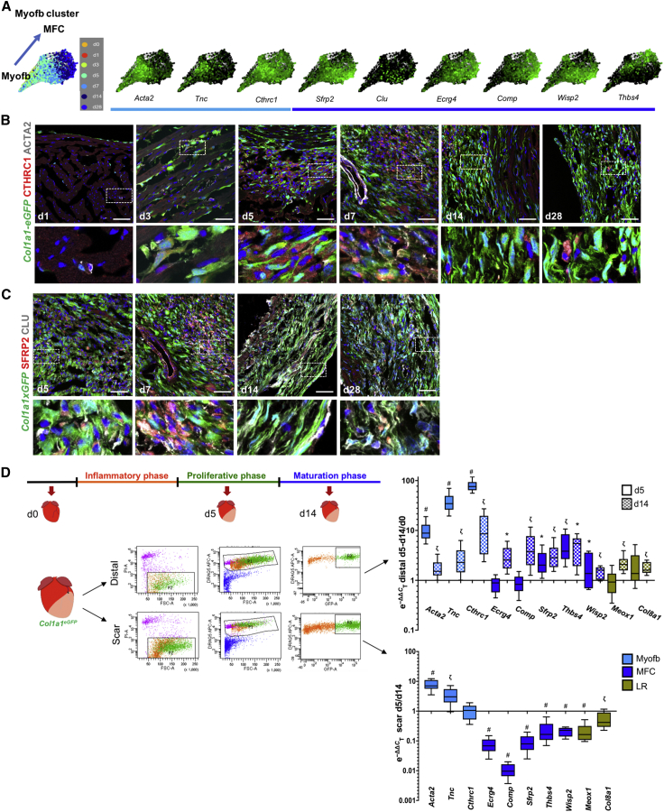
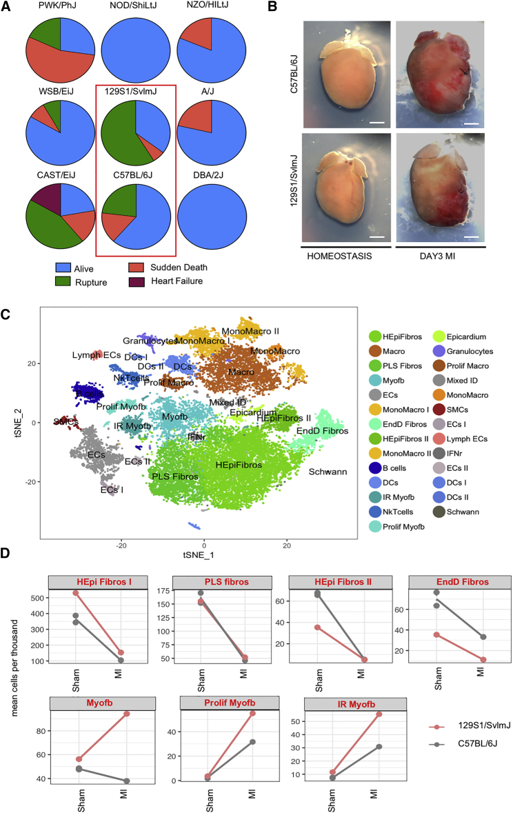
Cardiac ischemia leads to the loss of myocardial tissue and the activation of a repair process that culminates in the formation of a scar whose structural characteristics dictate propensity to favorable healing or detrimental cardiac wall rupture. To elucidate the cellular processes underlying scar formation, here we perform unbiased single-cell mRNA sequencing of interstitial cells isolated from infarcted mouse hearts carrying a genetic tracer that labels epicardial-derived cells. Sixteen interstitial cell clusters are revealed, five of which were of epicardial origin. Focusing on stromal cells, we define 11 sub-clusters, including diverse cell states of epicardial- and endocardial-derived fibroblasts. Comparing transcript profiles from post-infarction hearts in C57BL/6J and 129S1/SvImJ inbred mice, which displays a marked divergence in the frequency of cardiac rupture, uncovers an early increase in activated myofibroblasts, enhanced collagen deposition, and persistent acute phase response in 129S1/SvImJ mouse hearts, defining a crucial time window of pathological remodeling that predicts disease outcome.

摘要

心肌缺血导致心肌组织丧失,并激活修复过程,最终形成瘢痕,其结构特征决定了有利于愈合还是有害于心脏壁破裂的倾向。为了阐明瘢痕形成的细胞过程,我们在这里对携带标记心外膜细胞遗传示踪剂的梗死小鼠心脏分离的间质细胞进行了无偏单细胞 mRNA 测序。揭示了 16 个间质细胞簇,其中 5 个来自心外膜。我们专注于基质细胞,定义了 11 个亚簇,包括心外膜和心内膜来源成纤维细胞的多种细胞状态。比较 C57BL/6J 和 129S1/SvImJ 近交系小鼠梗死后心脏的转录谱,后者心脏破裂的频率明显不同,揭示了激活的肌成纤维细胞的早期增加、胶原沉积增强和 129S1/SvImJ 小鼠心脏中持续的急性期反应,定义了预测疾病结局的关键病理重塑时间窗口。

https://cdn.ncbi.nlm.nih.gov/pmc/blobs/887a/7059115/922cdd5ae15c/gr7.jpg
https://cdn.ncbi.nlm.nih.gov/pmc/blobs/887a/7059115/4681f0fd10fd/fx1.jpg
https://cdn.ncbi.nlm.nih.gov/pmc/blobs/887a/7059115/6eeca2af97d9/gr1.jpg
https://cdn.ncbi.nlm.nih.gov/pmc/blobs/887a/7059115/194614dfdc2f/gr2.jpg
https://cdn.ncbi.nlm.nih.gov/pmc/blobs/887a/7059115/2d36aa7bec1a/gr3.jpg
https://cdn.ncbi.nlm.nih.gov/pmc/blobs/887a/7059115/26f1388079f1/gr4.jpg
https://cdn.ncbi.nlm.nih.gov/pmc/blobs/887a/7059115/cdadaa82ce04/gr5.jpg
https://cdn.ncbi.nlm.nih.gov/pmc/blobs/887a/7059115/c87410801fca/gr6.jpg
https://cdn.ncbi.nlm.nih.gov/pmc/blobs/887a/7059115/922cdd5ae15c/gr7.jpg

相似文献

1
Dynamic Interstitial Cell Response during Myocardial Infarction Predicts Resilience to Rupture in Genetically Diverse Mice.心肌梗死后的动态间质细胞反应可预测遗传多样性小鼠对破裂的抵抗力。
Cell Rep. 2020 Mar 3;30(9):3149-3163.e6. doi: 10.1016/j.celrep.2020.02.008.
2
MK5 haplodeficiency decreases collagen deposition and scar size during post-myocardial infarction wound repair.MK5单倍体不足在心肌梗死后伤口修复过程中会减少胶原蛋白沉积和瘢痕大小。
Am J Physiol Heart Circ Physiol. 2019 Jun 1;316(6):H1281-H1296. doi: 10.1152/ajpheart.00532.2017. Epub 2019 Mar 22.
3
Novel technique for the simultaneous isolation of cardiac fibroblasts and epicardial stromal cells from the infarcted murine heart.从梗死的鼠心中同时分离心脏成纤维细胞和心外膜基质细胞的新方法。
Cardiovasc Res. 2020 Apr 1;116(5):1047-1058. doi: 10.1093/cvr/cvz193.
4
Reduced scar maturation and contractility lead to exaggerated left ventricular dilation after myocardial infarction in mice lacking AMPKα1.在缺乏AMPKα1的小鼠中,瘢痕成熟和收缩性降低导致心肌梗死后左心室扩张加剧。
J Mol Cell Cardiol. 2014 Sep;74:32-43. doi: 10.1016/j.yjmcc.2014.04.018. Epub 2014 May 5.
5
Akt-dependent Girdin phosphorylation regulates repair processes after acute myocardial infarction.Akt 依赖的 Girdin 磷酸化调节急性心肌梗死后的修复过程。
J Mol Cell Cardiol. 2015 Nov;88:55-63. doi: 10.1016/j.yjmcc.2015.09.012. Epub 2015 Sep 21.
6
Myocardial Infarction Induces Cardiac Fibroblast Transformation within Injured and Noninjured Regions of the Mouse Heart.心肌梗死诱导小鼠心脏损伤和非损伤区域的心肌成纤维细胞转化。
J Proteome Res. 2021 May 7;20(5):2867-2881. doi: 10.1021/acs.jproteome.1c00098. Epub 2021 Mar 31.
7
Interacting resident epicardium-derived fibroblasts and recruited bone marrow cells form myocardial infarction scar.心肌梗死后瘢痕由相互作用的心肌内源性成纤维细胞和募集的骨髓细胞形成。
J Am Coll Cardiol. 2015 May 19;65(19):2057-66. doi: 10.1016/j.jacc.2015.03.520.
8
Single-Cell RNA Sequencing Analysis Reveals a Crucial Role for CTHRC1 (Collagen Triple Helix Repeat Containing 1) Cardiac Fibroblasts After Myocardial Infarction.单细胞RNA测序分析揭示了CTHRC1(含胶原蛋白三螺旋重复序列1)在心肌梗死后心脏成纤维细胞中的关键作用。
Circulation. 2020 Nov 10;142(19):1831-1847. doi: 10.1161/CIRCULATIONAHA.119.044557. Epub 2020 Sep 25.
9
Properties and Functions of Fibroblasts and Myofibroblasts in Myocardial Infarction.心肌梗死中成纤维细胞和肌成纤维细胞的特性和功能。
Cells. 2022 Apr 20;11(9):1386. doi: 10.3390/cells11091386.
10
The role of cardiac fibroblasts in post-myocardial heart tissue repair.心脏成纤维细胞在心肌梗死后心脏组织修复中的作用。
Exp Mol Pathol. 2016 Oct;101(2):231-240. doi: 10.1016/j.yexmp.2016.09.002. Epub 2016 Sep 9.

引用本文的文献

1
SMAD7: riding on fibrosis-limiting routes and beyond.SMAD7:在限制纤维化的途径上前行及其他作用
EMBO Mol Med. 2025 Jul 31. doi: 10.1038/s44321-025-00283-7.
2
Immunoregulatory Endothelial Cells Interact With T Cells After Myocardial Infarction.免疫调节性内皮细胞在心肌梗死后与T细胞相互作用。
Circ Res. 2025 Aug 29;137(6):866-879. doi: 10.1161/CIRCRESAHA.125.326145. Epub 2025 Jul 30.
3
Temporal dynamics of the multi-omic response reveals the modulation of macrophage subsets post-myocardial infarction.多组学反应的时间动态揭示了心肌梗死后巨噬细胞亚群的调节。

本文引用的文献

1
Fast, sensitive and accurate integration of single-cell data with Harmony.利用 Harmony 实现单细胞数据的快速、灵敏和精确整合。
Nat Methods. 2019 Dec;16(12):1289-1296. doi: 10.1038/s41592-019-0619-0. Epub 2019 Nov 18.
2
DoubletFinder: Doublet Detection in Single-Cell RNA Sequencing Data Using Artificial Nearest Neighbors.DoubletFinder:基于人工最近邻算法检测单细胞 RNA 测序数据中的双细胞。
Cell Syst. 2019 Apr 24;8(4):329-337.e4. doi: 10.1016/j.cels.2019.03.003. Epub 2019 Apr 3.
3
Single-cell expression profiling reveals dynamic flux of cardiac stromal, vascular and immune cells in health and injury.
J Transl Med. 2025 Jul 10;23(1):777. doi: 10.1186/s12967-025-06726-6.
4
Emerging Epigenetic Therapies for the Treatment of Cardiac Fibrosis.用于治疗心脏纤维化的新兴表观遗传疗法。
Biomedicines. 2025 May 11;13(5):1170. doi: 10.3390/biomedicines13051170.
5
A cell type-specific expression atlas of small and total RNA in the heart after myocardial infarction.心肌梗死后心脏中小RNA和总RNA的细胞类型特异性表达图谱。
Sci Data. 2025 May 19;12(1):816. doi: 10.1038/s41597-025-05061-1.
6
Elucidating the preventive and therapeutic effects of cardiac telocytes paracrine microRNAs on ischemic heart disease.阐明心脏间质细胞旁分泌微小RNA对缺血性心脏病的预防和治疗作用。
Front Cardiovasc Med. 2025 Apr 1;12:1540051. doi: 10.3389/fcvm.2025.1540051. eCollection 2025.
7
Inhibiting the Histone Demethylase Kdm4a Restrains Cardiac Fibrosis After Myocardial Infarction by Promoting Autophagy in Premature Senescent Fibroblasts.抑制组蛋白去甲基化酶Kdm4a通过促进早衰成纤维细胞自噬来抑制心肌梗死后的心脏纤维化。
Adv Sci (Weinh). 2025 Jun;12(21):e2414830. doi: 10.1002/advs.202414830. Epub 2025 Apr 15.
8
Dynamic molecular atlas of cardiac fibrosis at single-cell resolution shows CD248 in cardiac fibroblasts orchestrates interactions with immune cells.单细胞分辨率下心脏纤维化的动态分子图谱显示,心脏成纤维细胞中的CD248协调与免疫细胞的相互作用。
Nat Cardiovasc Res. 2025 Apr;4(4):380-396. doi: 10.1038/s44161-025-00617-1. Epub 2025 Mar 27.
9
CD248-targeted BBIR-T cell therapy against late-activated fibroblasts in cardiac repair after myocardial infarction.靶向CD248的BBIR-T细胞疗法用于心肌梗死后心脏修复中晚期激活的成纤维细胞的治疗
Nat Commun. 2025 Mar 27;16(1):2895. doi: 10.1038/s41467-025-56703-2.
10
Macrophage-derived SPP1 exacerbate myocardial injury by interacting with fibroblasts in viral myocarditis.巨噬细胞衍生的SPP1通过与病毒性心肌炎中的成纤维细胞相互作用加剧心肌损伤。
Biol Direct. 2025 Mar 14;20(1):30. doi: 10.1186/s13062-025-00621-2.
单细胞表达谱分析揭示了心脏基质、血管和免疫细胞在健康和损伤中的动态变化。
Elife. 2019 Mar 26;8:e43882. doi: 10.7554/eLife.43882.
4
Variable outcomes of human heart attack recapitulated in genetically diverse mice.基因多样的小鼠再现了人类心脏病发作的不同结果。
NPJ Regen Med. 2019 Mar 4;4:5. doi: 10.1038/s41536-019-0067-6. eCollection 2019.
5
Regenerating the field of cardiovascular cell therapy.心血管细胞治疗领域的再生。
Nat Biotechnol. 2019 Mar;37(3):232-237. doi: 10.1038/s41587-019-0042-1. Epub 2019 Feb 18.
6
Fourth Universal Definition of Myocardial Infarction (2018).心肌梗死的第四次全球定义(2018年)。
Circulation. 2018 Nov 13;138(20):e618-e651. doi: 10.1161/CIR.0000000000000617.
7
Fate Mapping of Sca1 Cardiac Progenitor Cells in the Adult Mouse Heart.成年小鼠心脏中Sca1心脏祖细胞的命运图谱
Circulation. 2018 Dec 18;138(25):2967-2969. doi: 10.1161/CIRCULATIONAHA.118.036210.
8
Profiling proliferative cells and their progeny in damaged murine hearts.在受损的小鼠心脏中分析增殖细胞及其后代。
Proc Natl Acad Sci U S A. 2018 Dec 26;115(52):E12245-E12254. doi: 10.1073/pnas.1805829115. Epub 2018 Dec 7.
9
PANTHER version 14: more genomes, a new PANTHER GO-slim and improvements in enrichment analysis tools.PANTHER 版本 14:更多基因组、一个新的 PANTHER GO-slim 和富集分析工具的改进。
Nucleic Acids Res. 2019 Jan 8;47(D1):D419-D426. doi: 10.1093/nar/gky1038.
10
Following hearts, one cell at a time: recent applications of single-cell RNA sequencing to the understanding of heart disease.逐一追踪细胞:单细胞 RNA 测序在心脏疾病研究中的最新应用。
Nat Commun. 2018 Oct 30;9(1):4434. doi: 10.1038/s41467-018-06894-8.