Department of Cognitive Neuroscience, Donders Institute for Brain, Cognition and Behaviour, Radboudumc, Nijmegen, The Netherlands.
Bionics Institute, East Melbourne, Victoria, Australia.
Invest Ophthalmol Vis Sci. 2023 Mar 1;64(3):1. doi: 10.1167/iovs.64.3.1.
Most eye-movement studies in patients with visual field defects have examined the strategies that patients use while exploring a visual scene, but they have not investigated saccade kinematics. In healthy vision, saccade trajectories follow the remarkably stereotyped "main sequence": saccade duration increases linearly with saccade amplitude; peak velocity also increases linearly for small amplitudes, but approaches a saturation limit for large amplitudes. Recent theories propose that these relationships reflect the brain's attempt to optimize vision when planning eye movements. Therefore, in patients with bilateral retinal damage, saccadic behavior might differ to optimize vision under the constraints imposed by the visual field defects.
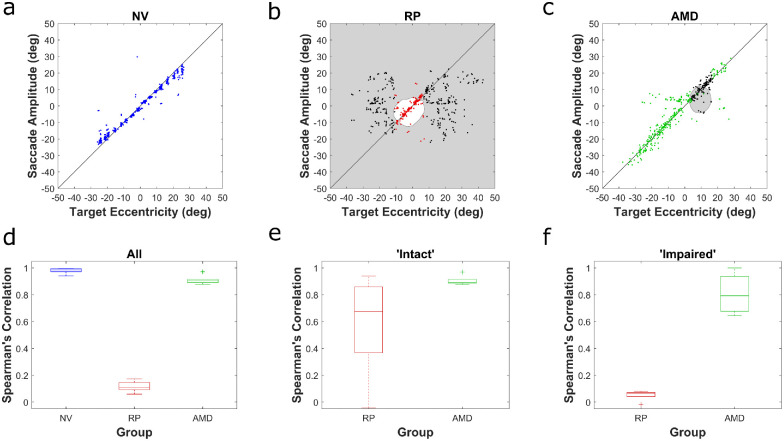
We compared saccadic behavior of patients with central vision loss, due to age-related macular degeneration (AMD), and patients with peripheral vision loss, due to retinitis pigmentosa (RP), to that of controls with normal vision (NV) using a horizontal saccade task.
Both patient groups demonstrated deficits in saccade reaction times and target localization behavior, as well as altered saccade kinematics. Saccades were generally slower and the shape of the velocity profiles were often atypical, especially in the patients with RP. In the patients with AMD, the changes were far less dramatic. For both groups, saccade kinematics were affected most when the target was in the subjects' blind field.
We conclude that defects of the central and peripheral retina have distinct effects on the saccade main sequence, and that visual inputs play an important role in planning the kinematics of a saccade.
大多数针对视野缺损患者的眼动研究都考察了患者在探索视觉场景时使用的策略,但并未研究眼跳运动学。在正常视力下,眼跳轨迹遵循显著的“标准序列”:眼跳持续时间随眼跳幅度线性增加;对于小幅度,峰值速度也线性增加,但对于大幅度,接近饱和极限。最近的理论提出,这些关系反映了大脑在规划眼球运动时试图优化视觉的尝试。因此,在双侧视网膜损伤的患者中,眼跳行为可能会有所不同,以在视野缺损所施加的限制下优化视觉。
我们使用水平眼跳任务比较了由于年龄相关性黄斑变性(AMD)而导致中央视力丧失的患者和由于色素性视网膜炎(RP)而导致周边视力丧失的患者与具有正常视力(NV)的对照组的眼跳行为。
两组患者的眼跳反应时间和目标定位行为以及眼跳运动学均存在缺陷。眼跳通常较慢,速度曲线的形状也常常不典型,尤其是在 RP 患者中。在 AMD 患者中,变化则不那么明显。对于两组患者,当目标处于受试者的盲区内时,眼跳运动学受到的影响最大。
我们得出结论,中央和周边视网膜的缺陷对视跳标准序列有不同的影响,视觉输入在规划眼跳运动学方面起着重要作用。